1 · Approach
Assess urgency and volume. Confirm the abnormality. Compare current kidney function with a baseline.
Electrolytes, CKD, AKI, stones, myeloma and AL amyloid in one staged teaching session.
Andrew Bland, MD, FACP, FAAP · October 21, 2026
This fictional summary-day capstone integrates electrolytes, CKD, AKI, stones, monoclonal proteins and nephrotic syndrome. Allow about 22 minutes. Discussion prompts are ungraded and supplement the original 30 cases.
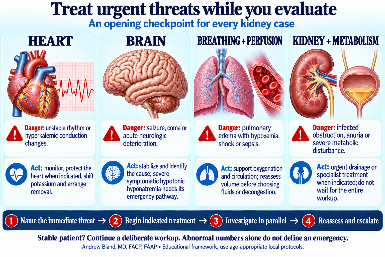
Name the threat, begin indicated treatment, investigate in parallel, then reassess.
Assess urgency and volume. Confirm the abnormality. Compare current kidney function with a baseline.
Use blood, urine and imaging together. A prior diagnosis does not explain every new abnormality.
Let PTH organize the calcium workup. Compare albumin with total urine protein.
Correct immediate threats, identify the lesion and clone, and reassess organ response.
Urgent-threat framework across the week · Opening lecture: kidney function and the skeleton key
A 64-year-old woman has one week of worsening thirst, frequent urination, constipation, poor intake and new slowed thinking. She also reports several weeks of back discomfort. She is not taking calcium supplements, high-dose vitamin D, lithium or a thiazide.
Her creatinine has been 1.2 mg/dL, with a reported eGFR near 50 mL/min/1.73 m² and UACR 65 mg/g, for more than six months. She passed a calcium oxalate stone two years ago. She took ibuprofen for her recent back pain.
She is afebrile, heart rate 108/min and BP 102/64 mmHg. Mucous membranes are dry; lungs are clear and there is no edema. She is slow to answer but follows commands.
| Test / finding | Result | Teaching reference / context |
|---|---|---|
| Total calcium / ionized calcium | 14.8 mg/dL / 1.85 mmol/L | 8.6–10.2 / 1.12–1.32 |
| Albumin / total serum protein | 3.6 / 9.2 g/dL | Albumin 3.5–5.0; protein 6.4–8.3 |
| Creatinine / BUN | 3.0 / 58 mg/dL | Creatinine was 1.2 one week ago |
| Na / K / bicarbonate | 146 / 3.2 / 25 mmol/L | Assess water balance, potassium deficit and acid–base status |
| Phosphate / alkaline phosphatase | 4.7 mg/dL / 96 U/L | 2.5–4.5 / 40–129; ALP is normal |
| Hemoglobin | 9.4 g/dL | Previously 12.1 g/dL |
| Urinalysis | 1+ protein; 0–2 RBC/hpf; no pyuria | Dipstick mainly detects albumin |
| Spot UPCR / UACR | 3.8 g/g / 180 mg/g | Total protein versus albumin: convert to matching units |
| Kidney/bladder ultrasound | Small nonobstructing stones; no hydronephrosis or bladder retention | Stones are present, but obstruction has not been demonstrated |
AKI evaluation lecture · Stone and obstruction case · Fluid choice and reassessment
| Requested marker | Patient result | Reference / interpretation task |
|---|---|---|
| Ca | 14.8 mg/dL; ionized 1.85 mmol/L | Confirmed hypercalcemia |
| PO₄ | 4.7 mg/dL | Mildly high during AKI; do not use phosphate alone to name the cause |
| Alk Phos | 96 U/L | Within the stated reference interval |
| 1,25(OH)₂ vitamin D | 12 pg/mL | 18–72; low |
| 25(OH) vitamin D | 31 ng/mL | Within this laboratory’s 20–50 interval |
| PTH | 6 pg/mL | 15–65; suppressed |
| PTHrP | <2.0 pmol/L | Not elevated; this assay’s upper limit is 4.2 |
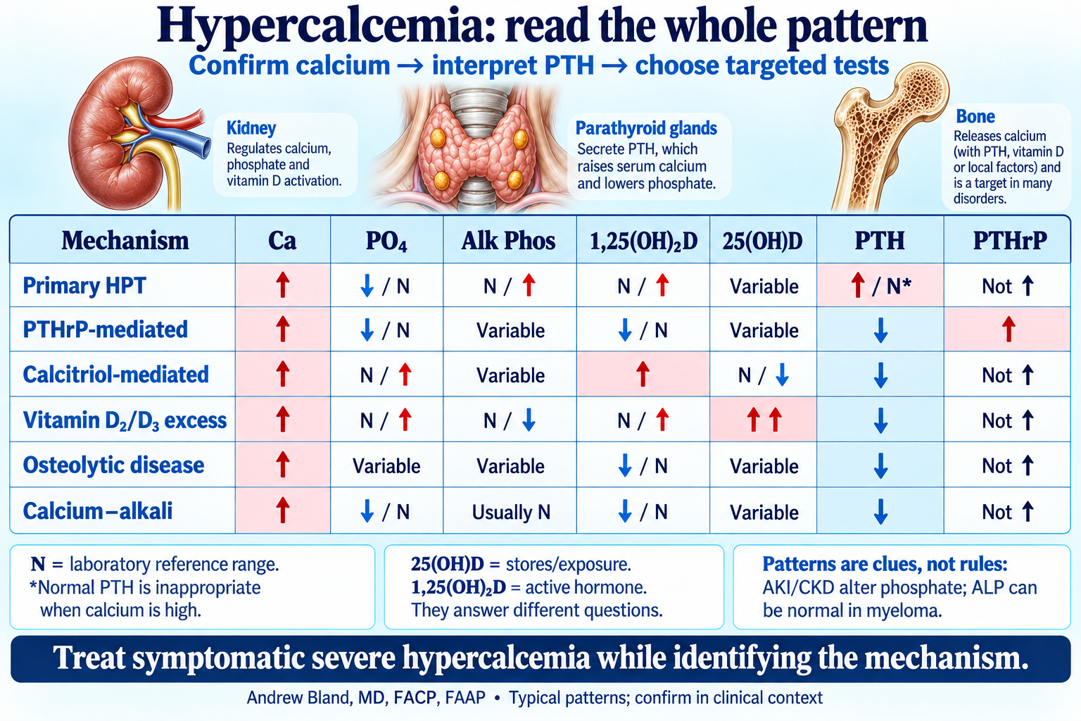
Ca, phosphate, alkaline phosphatase, both vitamin D metabolites, PTH and PTHrP.
| Mechanism | Ca | PO₄ | Alk Phos | 1,25(OH)₂D | 25(OH)D | PTH | PTHrP | Interpretation |
|---|---|---|---|---|---|---|---|---|
| Primary hyperparathyroidism | ↑ | ↓ or N | N or ↑ | N or ↑ | Variable | ↑ or inappropriately N | Not elevated | PTH should be suppressed by high calcium; check urine calcium in the appropriate setting. |
| PTHrP-mediated malignancy | ↑ | ↓ or N | Variable | ↓ or N | Variable | ↓ | ↑ | PTHrP mimics some PTH effects. An abnormal SPEP is not required. |
| Calcitriol-mediated disease | ↑ | N or ↑ | Variable | ↑ | N or ↓ | ↓ | Usually not elevated | Consider lymphoma or granulomatous disease; the lab pattern does not identify which. |
| Vitamin D₂/D₃ excess | ↑ | N or ↑ | N or ↓ | N or ↑ | Markedly ↑ | ↓ | Not elevated | Excess nutritional vitamin D raises 25(OH)D; active-vitamin-D medications are a different exposure. |
| Osteolytic malignancy / myeloma | ↑ | Variable | Variable; often N in myeloma | ↓ or N | Variable | ↓ | Usually not elevated | ALP can remain normal despite lytic lesions; evaluate monoclonal protein when indicated. |
| Calcium–alkali syndrome | ↑ | ↓ or N; affected by kidney function/intake | Usually N | ↓ or N | Variable | ↓ | Not elevated | Calcium/alkali exposure, alkalosis and AKI; medication history matters. |
| Tertiary hyperparathyroidism | ↑ | Often ↑ in advanced CKD; may fall after transplant | Variable | Variable; often low in advanced CKD | Variable | Markedly ↑ | Not elevated | Longstanding secondary HPT becomes autonomous; CKD alone does not prove this diagnosis. |
| Familial hypocalciuric hypercalcemia | Usually mildly ↑ | N or ↓ | Usually N | Variable | Variable | N or mildly ↑ | Not elevated | Lifelong/familial pattern and relative hypocalciuria; CKD, low vitamin D and thiazides confound urine calcium. |
Full hypercalcemia lecture and laboratory chart · Revisit Case 12: the renal mass and PTHrP pathway
Serum immunofixation identifies an IgG-kappa monoclonal protein; urine immunofixation identifies monoclonal kappa light chains. Serum free kappa is 2,400 mg/L and lambda 18 mg/L. Directed imaging shows lytic lesions. Bone marrow examination later shows 30% clonal plasma cells. These findings establish multiple myeloma with attributable myeloma-defining disease; a positive protein screen alone would not.
Kidney synthesis: volume depletion and hypercalcemia can reduce filtration, NSAID exposure can worsen perfusion, and the marked light-chain burden with mostly nonalbumin proteinuria raises concern for cast nephropathy. Cast injury is strongly suspected, not proven by the dipstick or FLC result alone. Nephrology decides whether tissue evaluation is needed, especially if the presentation or course is atypical.
| Time / goal | Teaching action |
|---|---|
| Now: stabilize | Monitored care; isotonic saline for volume depletion with repeated reassessment. Follow calcium, potassium, magnesium, phosphate, urine output and respiratory status. |
| Bridge: lower calcium | Calcitonin can provide a relatively rapid bridge; limit treatment to 48–72 hours because of tachyphylaxis. |
| Sustain: control hypercalcemia | For malignancy-associated hypercalcemia, select an IV bisphosphonate or denosumab with attention to kidney function and later hypocalcemia risk. These are not instant substitutes for stabilization. |
| Treat the cause | Urgent hematology/nephrology coordination for effective myeloma-directed treatment. Lowering calcium alone does not stop light-chain production. |
| Escalate and reassess | Dialysis is considered for refractory severe problems or when the clinical situation limits other treatment; neither creatinine nor FLC concentration alone is an automatic trigger. Routine loop diuretics are not used to force calcium excretion in a volume-depleted patient. |
Full student guide: monoclonal gammopathy and kidney disease
Compare a second fictional patient: progressive edema, serum albumin 2.1 g/dL, creatinine 1.6 mg/dL, calcium 9.4 mg/dL, UPCR 6.0 g/g and UACR 5,200 mg/g. Monoclonal studies identify a small lambda-producing clone. There are no lytic lesions.
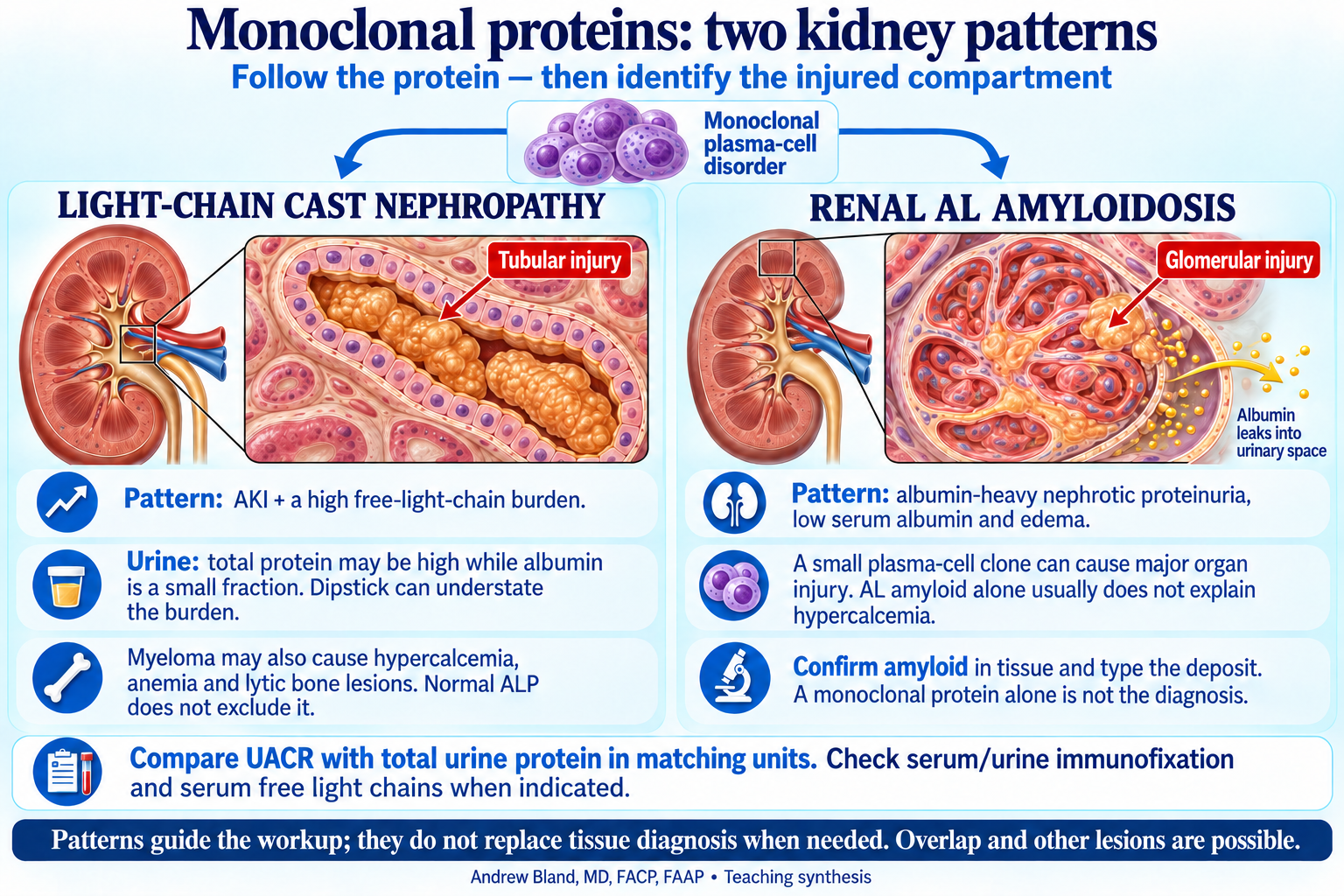
Tubular light-chain cast injury versus glomerular AL amyloid deposition.
Light-chain deposition disease (LCDD) has nonamyloid, nonfibrillar deposits and may involve glomerular, tubular and vascular basement membranes. It is another possible cause of proteinuria and kidney dysfunction. Congo-red negativity alone does not establish LCDD; light microscopy, immunofluorescence and electron microscopy define the lesion.
MGRS describes kidney injury attributable to a nephrotoxic monoclonal immunoglobulin from a clone that does not otherwise meet established hematologic treatment criteria. It is not a fixed marrow-percentage rule or a synonym for every kidney problem in a patient with an M-protein. Myeloma-associated cast nephropathy belongs to myeloma-related kidney disease.
Tests are complementary: SPEP/UPEP, immunofixation and serum FLC answer related questions. CKD affects both absolute FLC levels and ratio interpretation; use assay-appropriate reference intervals. A normal ratio does not by itself exclude AL or every MGRS lesion. MASS-FIX may identify a circulating monoclonal protein where available; it does not replace typing amyloid deposits in tissue.
Monoclonal kidney disease student guide · Amyloidosis and myeloma student guide · Edema lecture and diagnostic pathway · Compare Case 24: two nephrotic presentations
| System | How it fits this case |
|---|---|
| Electrolytes | Hypercalcemia explains several symptoms and can impair concentrating ability. Polyuria and poor intake contribute to water and potassium deficits. Phosphate must be read in the context of AKI. |
| CKD | Documented chronic G3a/A2 disease establishes reduced reserve. It does not explain the new creatinine rise or newly heavy nonalbumin protein excretion. |
| AKI | Volume depletion, hypercalcemic effects, NSAID exposure and possible cast injury can coexist. An unstable creatinine makes routine eGFR a poor estimate of current filtration. |
| Stones | Hypercalciuria can promote calcium stones, but an old stone does not establish the present calcium mechanism. Nonobstructing stones do not explain postrenal AKI; obstruction must be assessed. |
| Urine protein | UPCR and UACR measure different things. Nonalbumin-heavy proteinuria points toward a tubular/overflow pattern; albumin-heavy proteinuria with edema and low albumin suggests nephrotic glomerular disease. |
| Myeloma versus AL | Myeloma can cause hypercalcemia, anemia, lytic lesions and cast injury. AL amyloid can cause nephrotic syndrome from a small clone; tissue confirmation and typing remain essential. |
| Treatment | Stabilize immediate threats, treat the cause and track recovery. A calcium improvement, a falling light-chain level and a kidney response are related but separate endpoints. |
Return to Day 5 · Bring the lecture to the bedside · Visual library: summary-day figures