Visual summary
Sediment findings are powerful localization clues when the specimen is reliable and the clinical pattern fits.
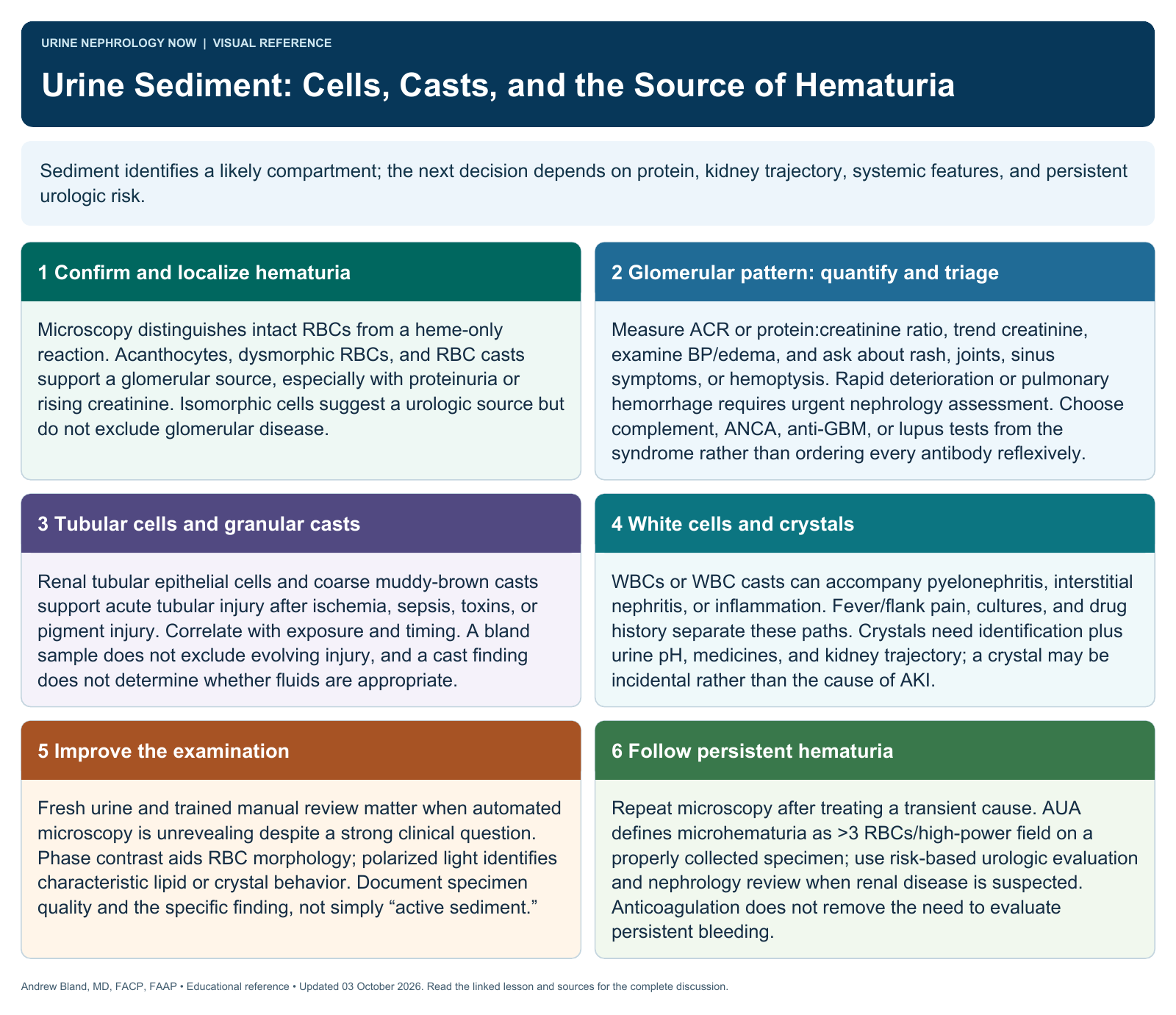
Text version
Formed elements localize injury
Urine microscopy can reveal red and white cells, tubular cells, casts, and crystals. These findings connect laboratory abnormalities to possible sites of injury, but collection, sample age, technique, and observer expertise affect reliability.
Recognize a glomerular pattern
Acanthocytes and red cell casts support glomerular bleeding, especially with proteinuria or declining kidney function. Isomorphic red cells can occur in urologic disease, but morphology is not an infallible exclusion test for glomerular pathology.
Assess the full sediment
Granular casts and tubular epithelial cells support tubular injury; white cell findings have inflammatory or infectious differentials. Crystals may be incidental or clinically important depending on type, pH, medication exposure, and the clinical setting.
Integrate and escalate
Combine microscopy with protein quantification, creatinine trend, BP, symptoms, and systemic findings. Rapid renal deterioration or a pulmonary–renal presentation warrants urgent specialist assessment; persistent hematuria also requires an appropriate urologic or nephrologic evaluation.
Use the right technique
Prompt processing and a trained examination improve interpretation. Phase contrast can help evaluate red cell morphology, and polarized light can assist with selected crystals or lipids. Automated reports may need manual review when the question is important.
Avoid absolute language
An absent cast does not exclude disease, and an unusual structure can be misidentified. Do not call every red cell cast universally pathognomonic or infer a precise diagnosis solely from a percent-dysmorphism threshold.
Continue learning
This graphic summarizes a topic. The full educational pages provide the broader discussion and references.