Visual summary
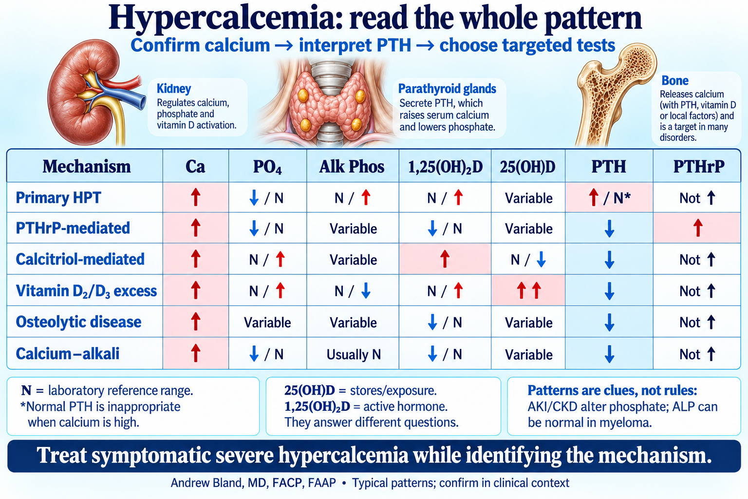
Ca, phosphate, alkaline phosphatase, both vitamin D metabolites, PTH and PTHrP.
Teaching text
Start with PTH in the context of calcium
A PTH inside its reference interval is not necessarily appropriate: high calcium should suppress it. A clearly suppressed PTH directs the workup toward PTH-independent causes. Use the assay and clinical context rather than a universal cutoff.
The vitamin D tests answer different questions
25-hydroxyvitamin D reflects stores and exposure. 1,25-dihydroxyvitamin D is the active hormone; an elevated value with suppressed PTH can suggest extrarenal production. A low 25-hydroxy level does not exclude calcitriol-driven hypercalcemia.
Phosphate and alkaline phosphatase are supporting clues
AKI and CKD can raise phosphate and blur a typical phosphaturic pattern. Total alkaline phosphatase includes liver and bone sources; normal ALP does not exclude myeloma or lytic bone disease. These tests help interpret a mechanism but cannot identify one alone.
The table is a pattern guide, not an order set
Not every patient needs all seven tests at once. Confirm calcium, assess symptoms and volume, interpret PTH, then choose PTHrP, vitamin D metabolites and monoclonal-protein testing as indicated. Mixed mechanisms and drug exposures can coexist.
Additional causes remain part of the differential
Thiazides can unmask primary hyperparathyroidism; lithium can shift calcium sensing. Vitamin A excess, immobilization, thyrotoxicosis, adrenal insufficiency and pediatric syndromes require the appropriate history and targeted tests. None has a single obligatory seven-test signature.
| Mechanism | Ca | PO₄ | Alk Phos | 1,25(OH)₂D | 25(OH)D | PTH | PTHrP | Interpretation |
|---|---|---|---|---|---|---|---|---|
| Primary hyperparathyroidism | ↑ | ↓ or N | N or ↑ | N or ↑ | Variable | ↑ or inappropriately N | Not elevated | PTH should be suppressed by high calcium; check urine calcium in the appropriate setting. |
| PTHrP-mediated malignancy | ↑ | ↓ or N | Variable | ↓ or N | Variable | ↓ | ↑ | PTHrP mimics some PTH effects. An abnormal SPEP is not required. |
| Calcitriol-mediated disease | ↑ | N or ↑ | Variable | ↑ | N or ↓ | ↓ | Usually not elevated | Consider lymphoma or granulomatous disease; the lab pattern does not identify which. |
| Vitamin D₂/D₃ excess | ↑ | N or ↑ | N or ↓ | N or ↑ | Markedly ↑ | ↓ | Not elevated | Excess nutritional vitamin D raises 25(OH)D; active-vitamin-D medications are a different exposure. |
| Osteolytic malignancy / myeloma | ↑ | Variable | Variable; often N in myeloma | ↓ or N | Variable | ↓ | Usually not elevated | ALP can remain normal despite lytic lesions; evaluate monoclonal protein when indicated. |
| Calcium–alkali syndrome | ↑ | ↓ or N; affected by kidney function/intake | Usually N | ↓ or N | Variable | ↓ | Not elevated | Calcium/alkali exposure, alkalosis and AKI; medication history matters. |
| Tertiary hyperparathyroidism | ↑ | Often ↑ in advanced CKD; may fall after transplant | Variable | Variable; often low in advanced CKD | Variable | Markedly ↑ | Not elevated | Longstanding secondary HPT becomes autonomous; CKD alone does not prove this diagnosis. |
| Familial hypocalciuric hypercalcemia | Usually mildly ↑ | N or ↓ | Usually N | Variable | Variable | N or mildly ↑ | Not elevated | Lifelong/familial pattern and relative hypocalciuria; CKD, low vitamin D and thiazides confound urine calcium. |
Continue learning
Read the connected teaching · Summary-day capstone · Bring the lecture to the bedside
Supporting evidence
- ARUP Laboratories: hypercalcemia testing algorithm
- Fifth International Workshop: primary hyperparathyroidism (2022)
- Endocrine Society: hypercalcemia of malignancy guideline
- Society for Endocrinology: emergency management of acute hypercalcaemia
- Alkaline phosphatase in myeloma and solid cancers with bone lesions
- Vitamin D intoxication and hypercalcemia