Visual summary
The first task is to characterize the lesion and the patient’s context, then select the least burdensome effective strategy.
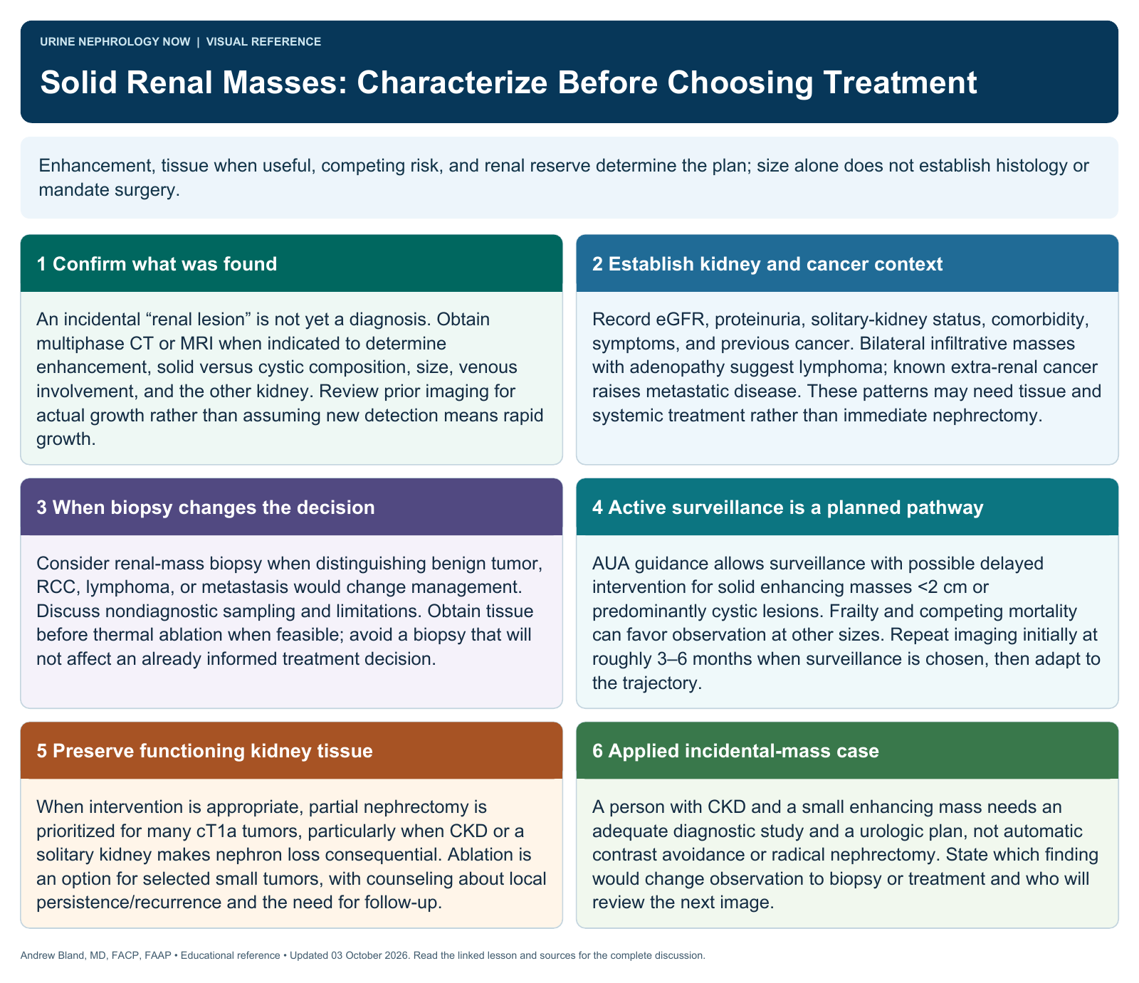
Text version
A renal mass has a differential
Renal cell carcinoma, benign tumors, lymphoma, and metastatic disease can produce renal masses. Imaging pattern, multiplicity, cancer history, and systemic findings affect the differential, but imaging does not always provide a definitive tissue diagnosis.
Recognize incidental and urgent presentations
Many lesions are found during unrelated imaging. Hematuria, pain, systemic symptoms, obstruction, or impaired renal function may change urgency. An incidental finding still needs clear characterization and a documented plan.
Choose informative imaging
Use an appropriate renal-mass protocol and assess whether contrast is needed to answer the question. Kidney function influences planning but should not automatically prevent an essential study; radiology and urology can help select the best approach.
Individualize management
Active surveillance, biopsy, ablation, nephron-sparing surgery, or systemic treatment may be appropriate in different settings. Preserve renal function when feasible while accounting for cancer risk, anatomy, comorbidity, treatment burden, and preferences.
Applied case: incidental mass
Case 19 integrates lesion characterization with kidney-risk assessment and multidisciplinary planning. Learners should explain how imaging will change the decision and when biopsy could distinguish primary renal cancer from lymphoma, metastasis, or a benign process.
Avoid size-only certainty
Size informs risk but cannot provide an exact metastatic probability for every lesion. A suspected lymphoma or metastatic lesion may need a different diagnostic and treatment pathway from a typical localized renal cell carcinoma.
Continue learning
This graphic summarizes a topic. The full educational pages provide the broader discussion and references.