Day 1: mechanisms behind the cases

Seventeen infographics for fluids, electrolytes, AKI and urine interpretation

Andrew Bland, MD, FACP, FAAP · October 20, 2026

Bring the lecture to the bedside — open the complete Day 1 teaching collection →

Use these figures alongside Cases 1–3 and their linked lectures. Each image opens at full size and has a text companion and download. The case files supply the patient data; the mechanisms explain the decisions.

Moving potassium is not removing potassium

HD began six weeks ago and urine output remains about 1 L/day. Trimethoprim can therefore impair residual renal potassium secretion; missed dialysis and a potassium salt substitute add risk. Urine volume does not quantify potassium clearance. Potassium 7.3 with a wide QRS and pulmonary edema requires calcium, shifting therapy and urgent HD. Lokelma can be given during the wait; a possible loop response must not delay the run.
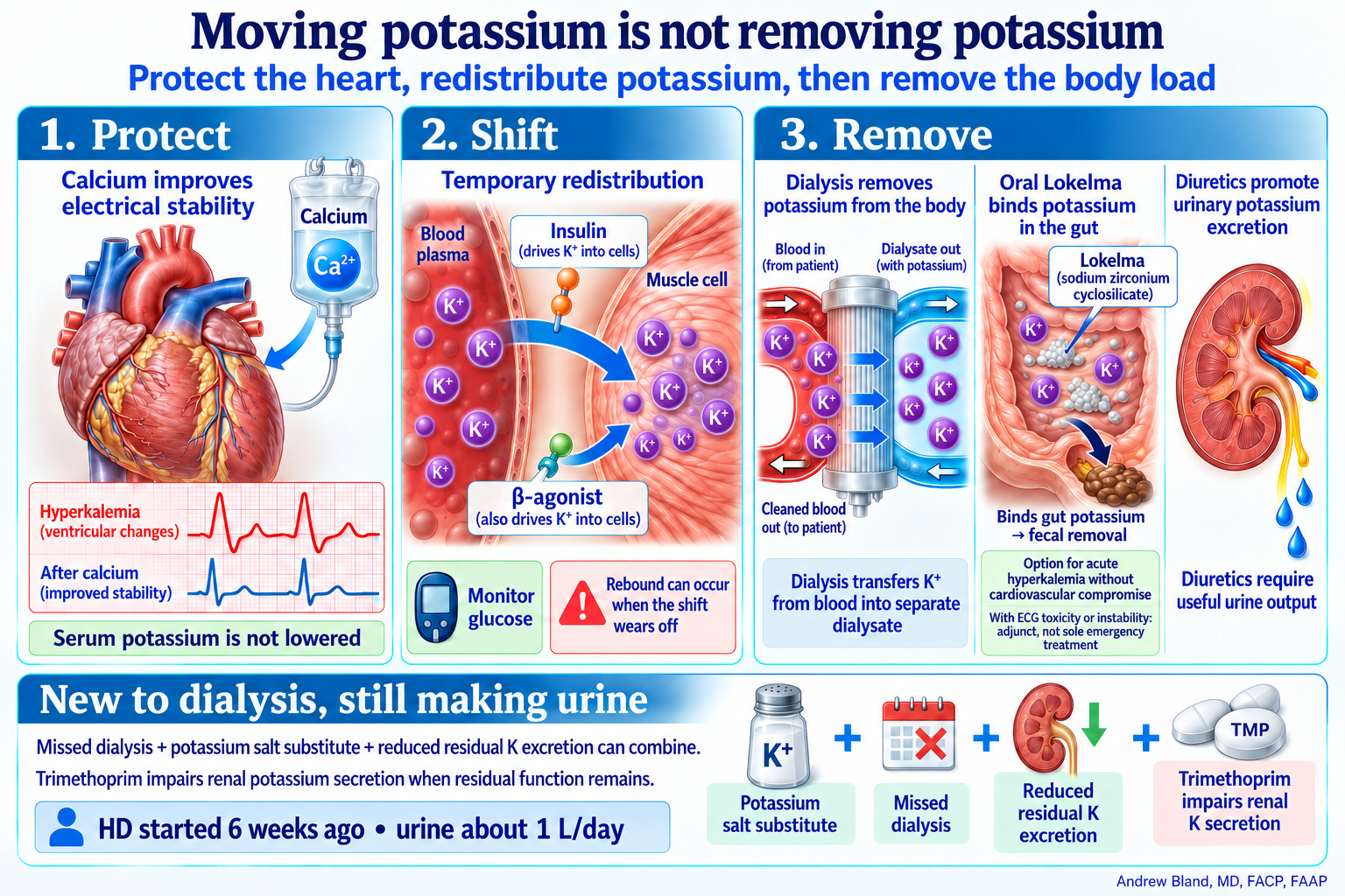
Calcium stabilizes the myocardium without lowering potassium. Insulin and beta agonists move potassium from blood into cells. Dialysis, gut binding with Lokelma, and useful urinary excretion remove potassium. Case 1 started HD six weeks ago and still makes about one liter of urine daily.
· Download PNG · Download Markdown · Read text and evidence

Protect the heart, redistribute potassium, then remove the body load

Read the serum, the urine, and the fluid

Normal saline contains 154 mEq/L sodium and has an osmolarity near 308 mOsm/L; 3% saline contains 513 mEq/L sodium and is about 1,026 mOsm/L. Do not compare sodium concentration directly with osmolality. Fluid osmolality above urine osmolality is a useful heuristic, not a universal requirement for sodium to rise. With persistent antidiuresis, sufficiently electrolyte-rich urine can excrete the infused salt in less water than was infused, leaving water behind (desalination). In volume depletion, saline may instead suppress ADH and release a water diuresis. Follow serial serum sodium, urine Na + K and urine volume.
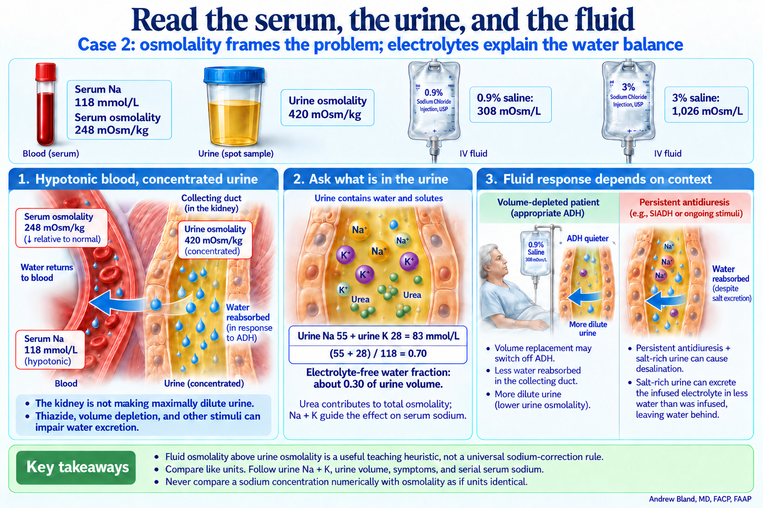
Case 2 has serum sodium 118, serum osmolality 248 and urine osmolality 420. Urine sodium 55 plus potassium 28 equals 83; dividing by serum sodium gives 0.70. Normal saline is 308 mOsm/L and 3% saline is 1,026 mOsm/L. Fluid response depends on urine electrolyte content, volume and changing ADH activity.
· Download PNG · Download Markdown · Read text and evidence

Case 2: osmolality frames the problem; electrolytes explain the water balance

Why water stays when sodium is low

Case 2 pairs hypotonic blood with concentrated urine in a patient taking a thiazide who is volume depleted and eating poorly. This is a water-handling problem with several contributors. A concentrated urine, even with a high urine sodium, does not independently establish SIAD.
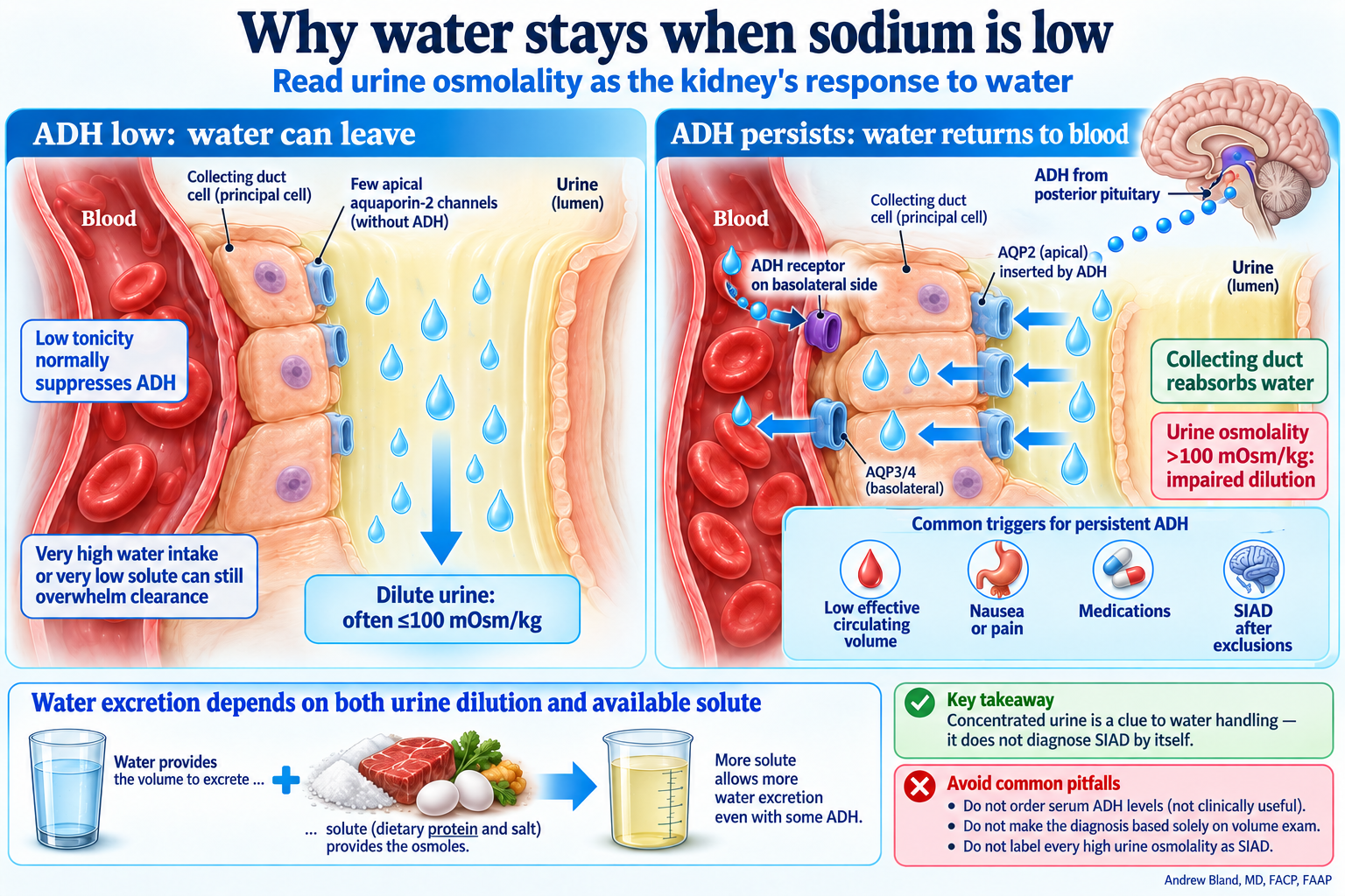
With low ADH, little water crosses the collecting-duct epithelium and dilute urine leaves the body. Persistent ADH signals the blood-side receptor and inserts apical aquaporins, permitting water movement from urine through cells into blood. Urine concentration reflects water handling but does not establish SIAD by itself.
· Download PNG · Download Markdown · Read text and evidence

Read urine osmolality as the kidney's response to water

One thiazide, several connected abnormalities

A diuretic can keep urine sodium and chloride high despite volume depletion. Those results do not independently prove SIAD or chloride-resistant alkalosis. Stop the thiazide, assess symptoms and volume, replace deficits and follow the response. Restored water excretion and potassium replacement can accelerate sodium correction.
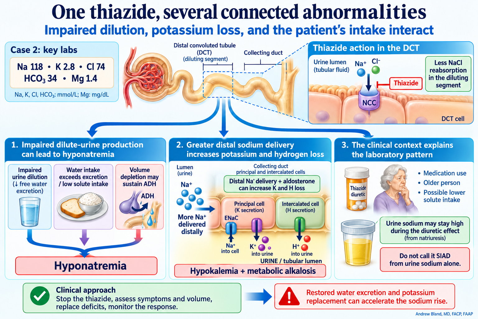
Thiazide inhibition of distal-convoluted-tubule NCC impairs urinary dilution. Water intake, low solute intake and volume-driven ADH can combine to lower sodium. Distal sodium delivery and aldosterone can increase potassium and hydrogen secretion, connecting hypokalemia and metabolic alkalosis.
· Download PNG · Download Markdown · Read text and evidence

Impaired dilution, potassium loss, and the patient's intake interact

When recovery makes sodium rise too fast

Escalate urgently to a monitored setting and nephrology; stop sodium-raising infusions and reassess all inputs, including potassium replacement. Specialist-directed desmopressin halts aquaresis and titrated free water permits controlled relowering, guided by frequent sodium checks and hourly urine output. In the fictional course, sodium returns to 126 by hour 18 and stays there through hour 24; no ODS develops. Rescue does not establish that the preceding overshoot was safe.
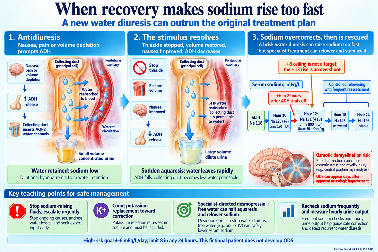
Case 2 sodium rises from 118 to 125 by hour 10, then to 131 at hour 12: six mEq/L in two hours and thirteen overall. Urine output reaches 800 mL/h with urine osmolality 80 after ADH falls. Specialist-directed rescue relowers sodium to 126 at hour 18 and holds it through hour 24. ODS can appear days after apparent improvement.
· Download PNG · Download Markdown · Read text and evidence

A new water diuresis can outrun the original treatment plan

Why potassium may not stay corrected

Address magnesium alongside potassium; do not delay urgent potassium replacement while waiting for magnesium to finish. Route, dose and monitoring depend on severity, symptoms and kidney function. Recheck both electrolytes and remember that potassium replacement contributes to sodium correction in this hyponatremic patient.
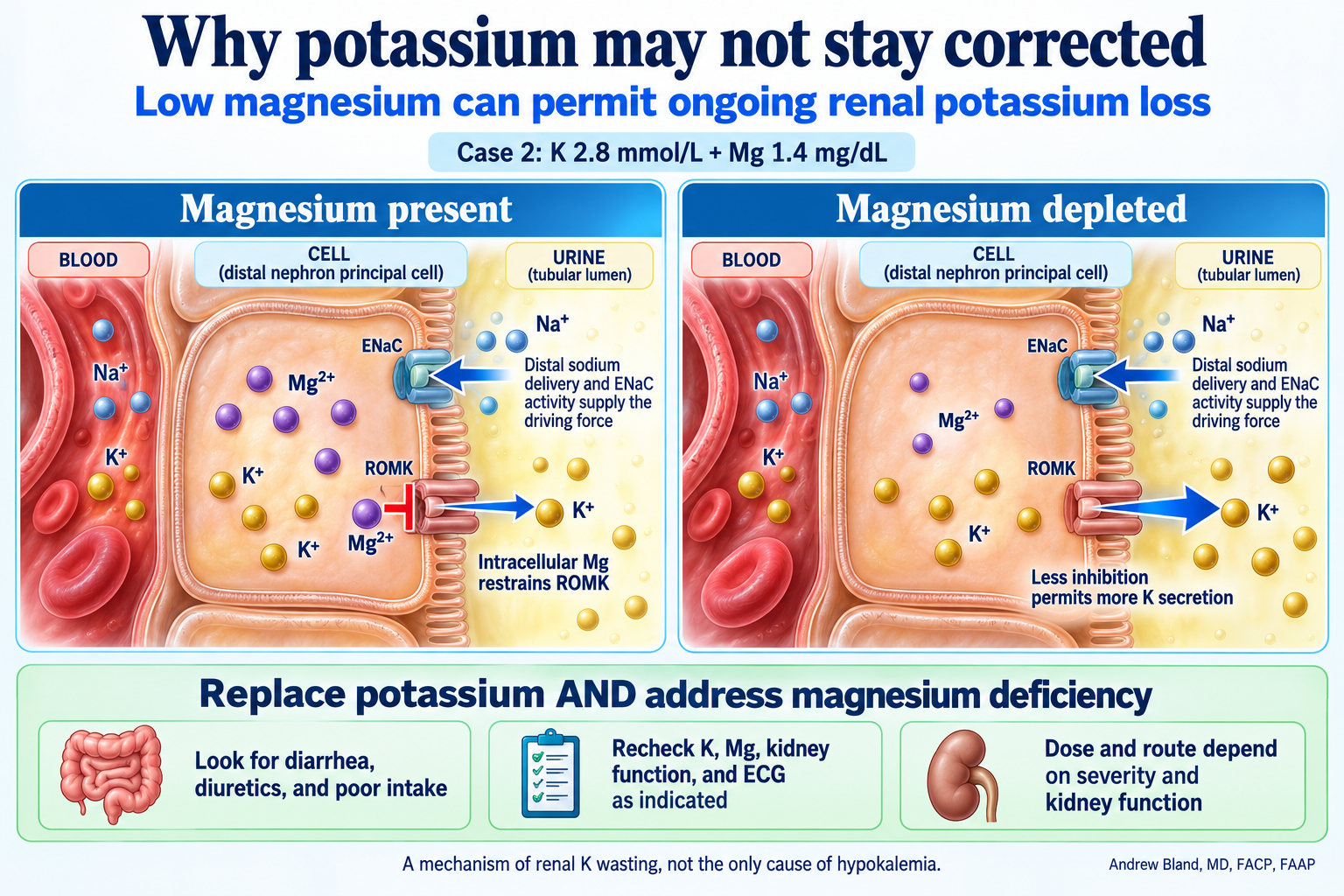
Intracellular magnesium normally restrains ROMK potassium secretion into urine. Magnesium depletion reduces that inhibition, allowing more potassium loss when distal sodium delivery and ENaC activity provide the driving force. Case 2 has potassium 2.8 and magnesium 1.4.
· Download PNG · Download Markdown · Read text and evidence

Low magnesium can permit ongoing renal potassium loss

Ketoacidosis can hide behind a modest glucose

Insulin stops ketone production; dextrose supports continued insulin when glucose is already modest or falls. Follow blood ketones, pH or bicarbonate, electrolytes and clinical status. Anion gap alone can mislead during recovery, especially after chloride-rich fluids. Hold the SGLT2 inhibitor and use the case's separate specialist review before any restart.
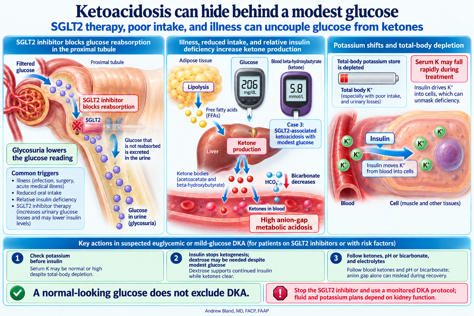
SGLT2 inhibition causes glycosuria while illness, poor intake and relative insulin deficiency can drive adipose lipolysis and liver ketone production. Case 3 glucose is 206 mg/dL and beta-hydroxybutyrate 5.8 mmol/L. Insulin moves potassium into cells, so potassium must be assessed before treatment and throughout recovery.
· Download PNG · Download Markdown · Read text and evidence

SGLT2 therapy, poor intake, and illness can uncouple glucose from ketones

Acute hyperkalemia: potassium and the ECG
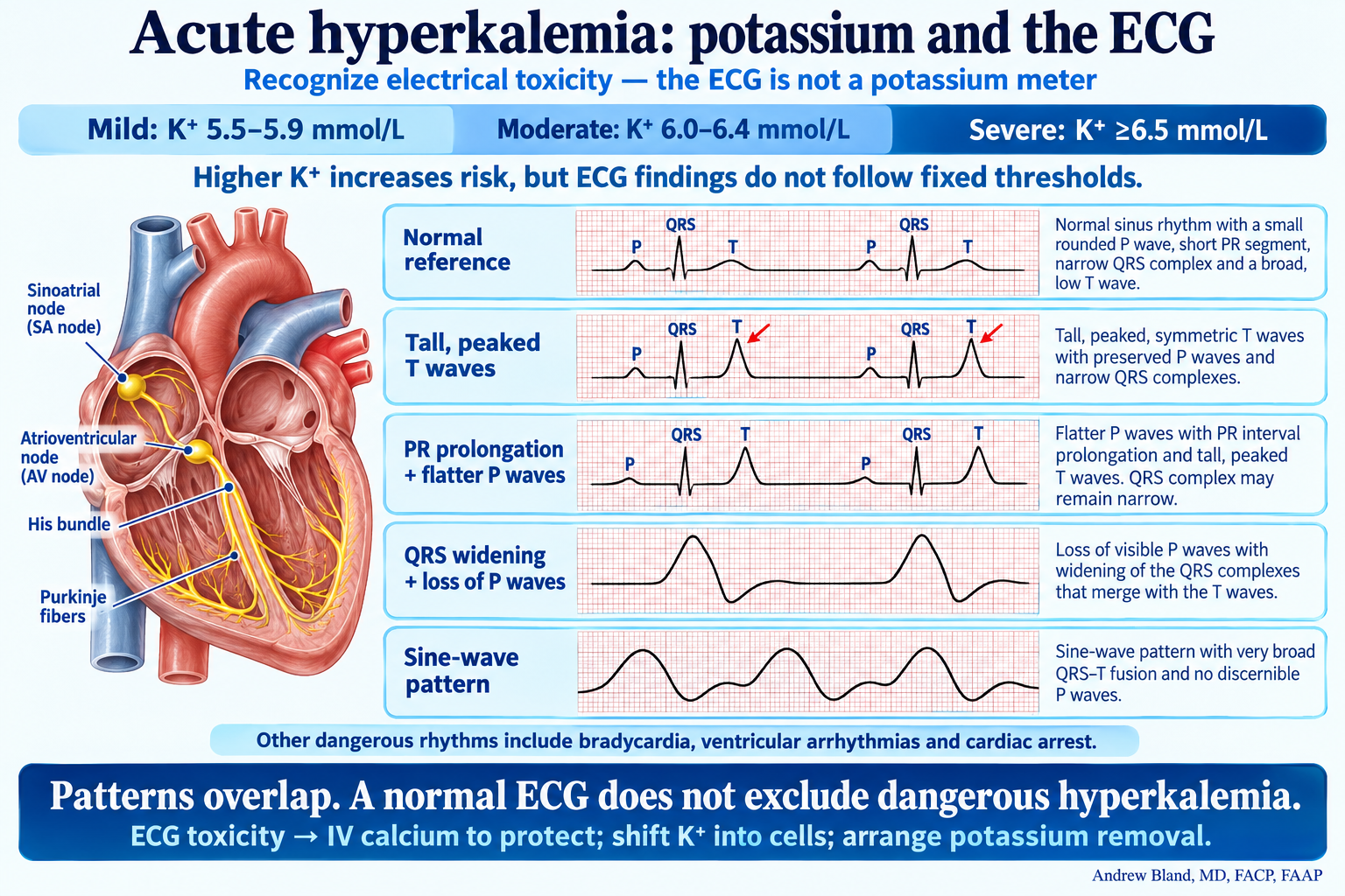
Potassium severity ranges are shown separately from ECG patterns: peaked T waves, PR prolongation, flatter or absent P waves, QRS widening and sine-wave fusion. A normal ECG does not exclude dangerous hyperkalemia.
· Download PNG · Download Markdown · Read text and evidence

Recognize electrical toxicity; the ECG does not measure potassium.

Open the complete teaching text and evidence →

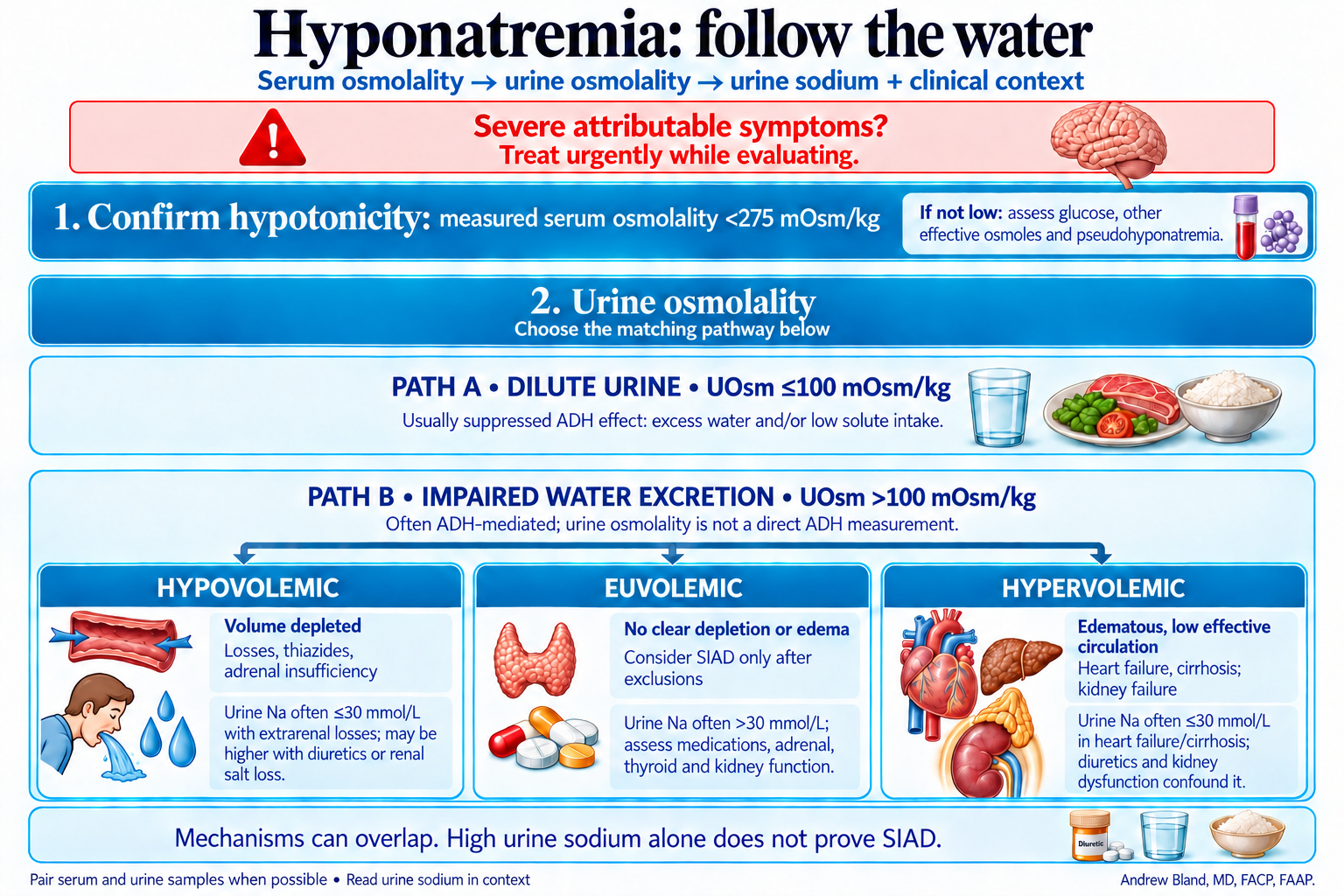
Hyponatremia: follow the water

Two alternative urine-osmolality pathways: dilute urine at or below 100 mOsm/kg, or impaired water excretion above 100. The latter separates into hypovolemic, euvolemic and hypervolemic swim lanes with contextual urine sodium interpretation.
· Download PNG · Download Markdown · Read text and evidence

Follow the original lecture lanes: tonicity, urine dilution, then volume and urine sodium.

Open the complete teaching text and evidence →

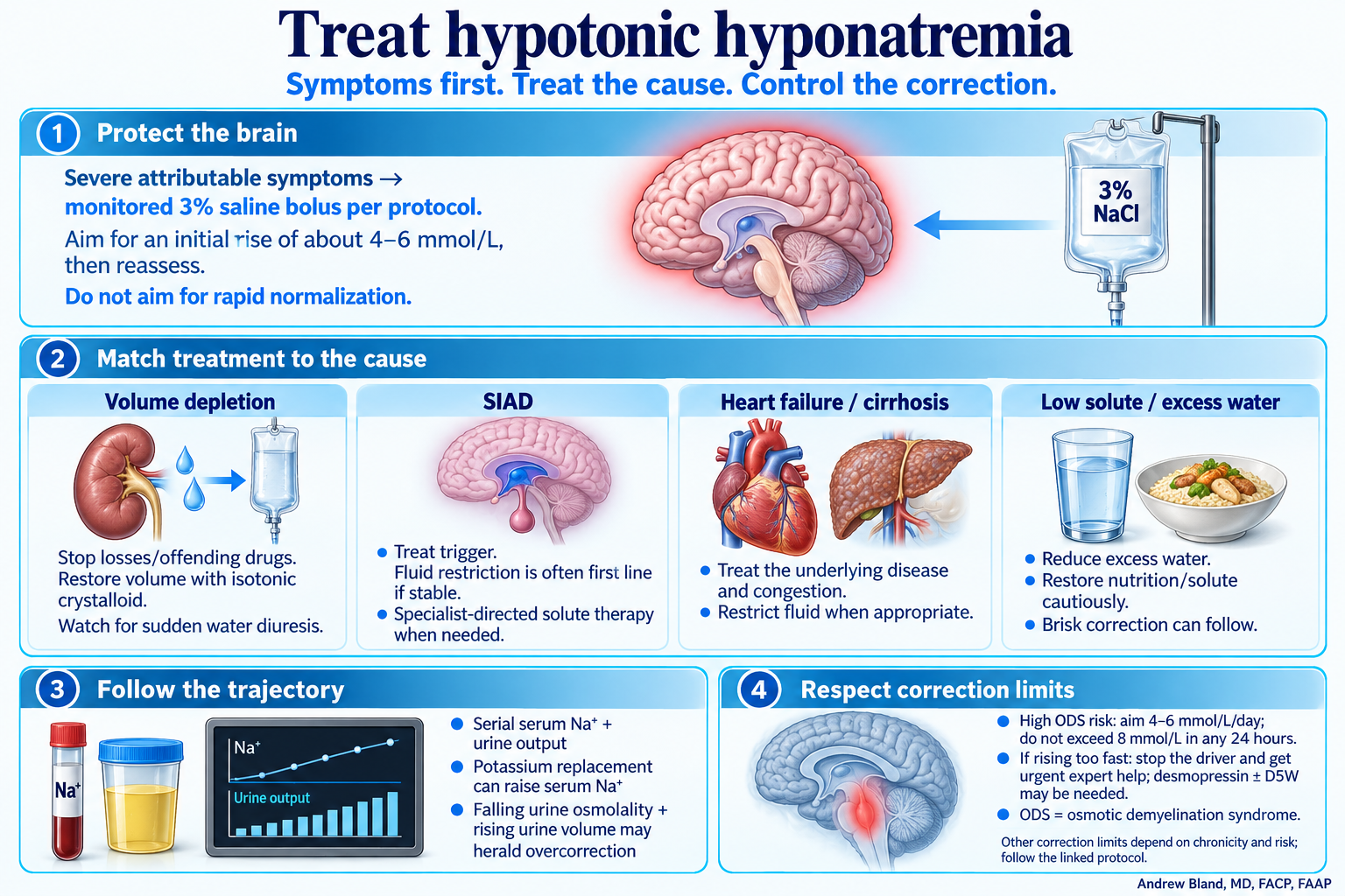
Treat hypotonic hyponatremia

Severe attributable symptoms prompt monitored hypertonic saline. Stable treatment follows volume depletion, SIAD, hypervolemic disease, or excess water/low solute pathways, with serial sodium and urine-output monitoring.
· Download PNG · Download Markdown · Read text and evidence

Symptoms first, cause-directed treatment, and control of the correction.

Open the complete teaching text and evidence →

Urine sediment: clues to the AKI mechanism
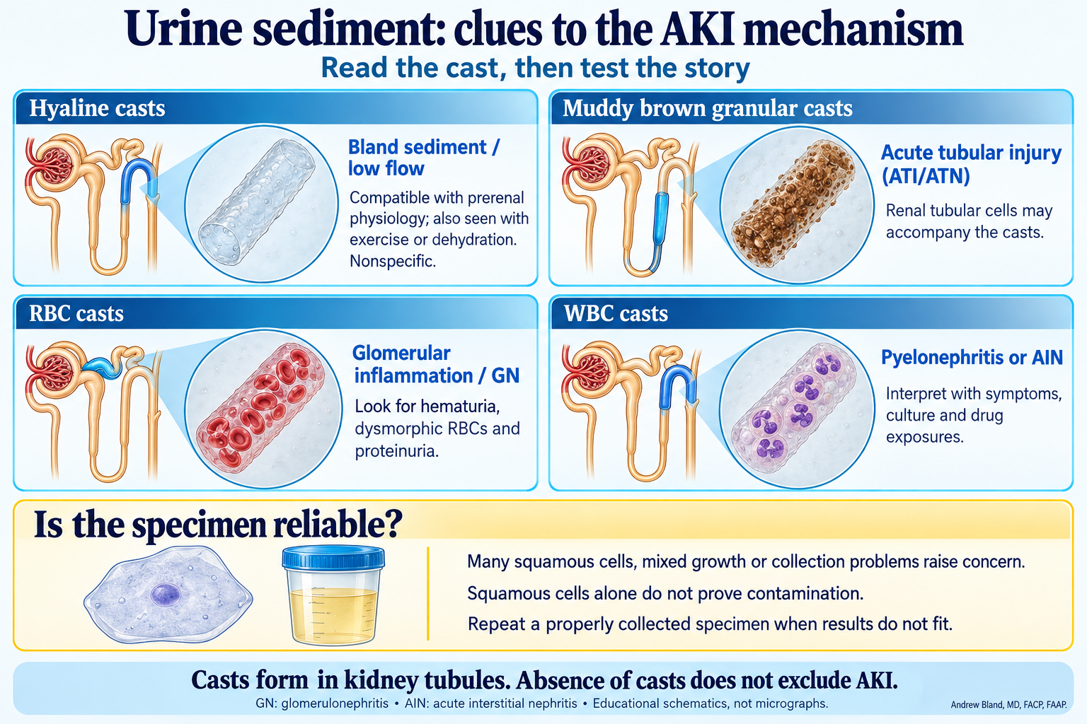
Schematic hyaline, muddy brown granular, red-cell and white-cell casts, with their common clinical associations and a separate section on squamous epithelial cells and possible specimen contamination.
· Download PNG · Download Markdown · Read text and evidence

Read the cast, then test the clinical story and specimen quality.

Open the complete teaching text and evidence →

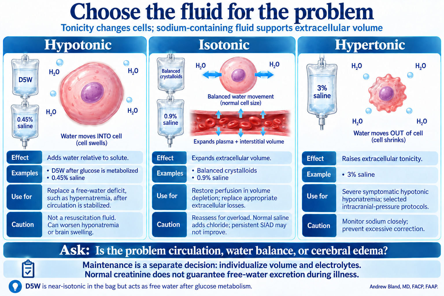
Choose the fluid for the problem

Hypotonic fluid adds water relative to solute; isotonic crystalloid expands plasma and interstitial volume; hypertonic saline raises extracellular tonicity. Maintenance fluid requires a separate individualized decision.
· Download PNG · Download Markdown · Read text and evidence

Separate circulation, water balance and cerebral edema from routine maintenance.

Open the complete teaching text and evidence →

Lactated Ringer’s versus normal saline
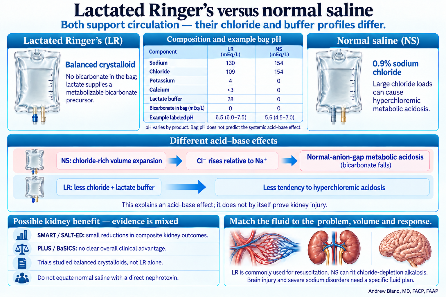
LR and saline electrolyte comparison. Chloride-rich saline can cause normal-anion-gap acidosis; LR has less chloride and a metabolizable buffer. Kidney-outcome trials of balanced crystalloids have mixed results.
· Download PNG · Download Markdown · Read text and evidence

Different chloride and buffer profiles; possible kidney benefit with mixed clinical evidence.

Open the complete teaching text and evidence →

Acute Kidney Injury: Recognize, Localize, Reassess
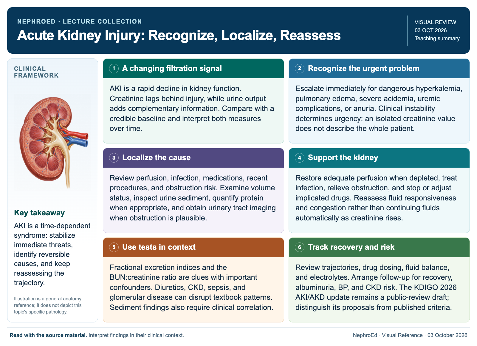
Existing AKI framework: recognize the creatinine or urine-output change, assess complications, distinguish impaired perfusion, intrinsic injury and obstruction, and reassess.
· Download PNG · Download Markdown · Read text and evidence

Prerenal, intrinsic and postrenal mechanisms can overlap. Reassess the response.

Open the complete teaching text and evidence →

Acute hyperkalemia: buy time, then remove potassium
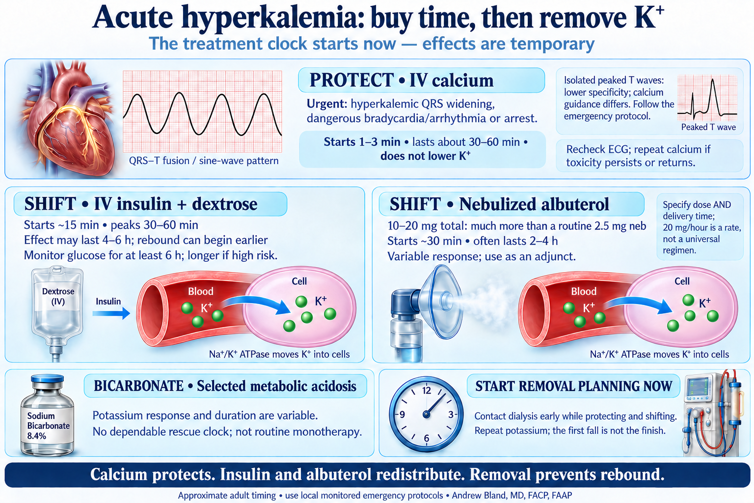
Treatment clocks: calcium begins within minutes and protects for about 30–60 minutes; insulin and albuterol temporarily shift potassium; bicarbonate has an inconsistent potassium response. Removal planning starts alongside these interventions.
· Download PNG · Download Markdown · Read text and evidence

Calcium protection and intracellular shifts have different clocks; start removal planning immediately.

Hyperkalemia: remove potassium while the clock runs
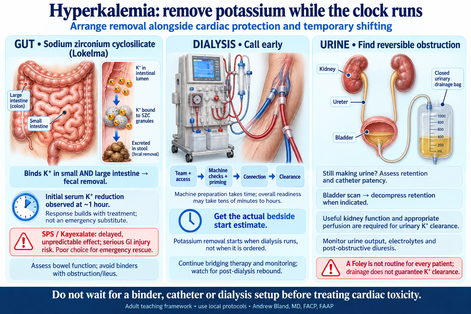
Three potassium-removal pathways: Lokelma binds potassium in small and large intestine, dialysis requires team/access/machine preparation before clearance begins, and catheterization relieves suspected urinary retention when indicated. Emergency cardiac treatment continues in parallel.
· Download PNG · Download Markdown · Read text and evidence

Gut, dialysis and useful urinary clearance are removal routes; none should delay cardiac stabilization.

Treat urgent threats while you evaluate
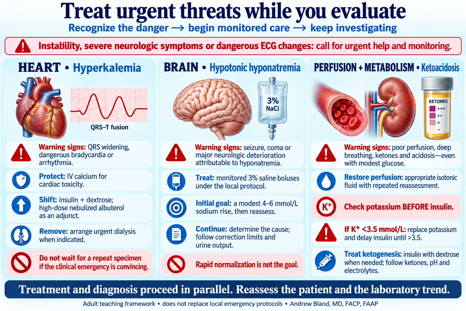
Three urgent pathways: calcium, shifting and removal for hyperkalemic cardiac toxicity; monitored hypertonic saline for severe symptoms attributable to hypotonic hyponatremia; perfusion support and potassium-aware insulin treatment for ketoacidosis.
· Download PNG · Download Markdown · Read text and evidence

Recognize danger, begin monitored care and keep investigating in parallel.