Start with the physiology. Turn the key. Follow the timeline.
Andrew Bland, MD, FACP, FAAP · October 2026
Start here · Before the first case
Why do we have kidneys? To keep the internal environment livable as the world around us changes. Begin with that job, then use blood work, urine, imaging, and the timeline to understand what has gone wrong.
01 · Begin with the purpose
Why have kidneys? Life out of water.
We live on land, but our cells still need a carefully controlled watery environment.
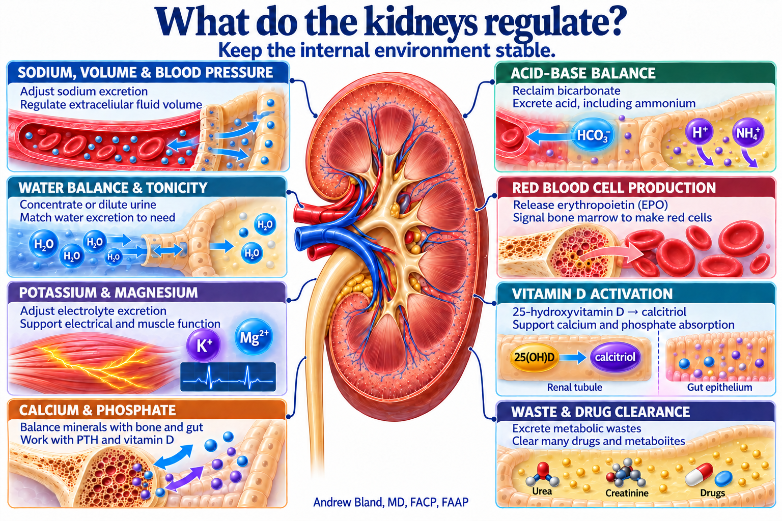
Kidneys help make life away from water possible: conserve water and salt when supplies are scarce, release excess when supplies are abundant, and remove wastes. They preserve the internal environment despite changing intake and losses.
A water-poor, salt-poor environment makes conservation essential. Modern salt excess challenges the same regulatory systems. Kidney disease becomes easier to understand when we ask which part of this balancing act has failed.
Start the discussion: If you could no longer adjust what leaves in the urine, what would happen after a day without water? After a salty meal?
Total-body sodium helps determine extracellular fluid volume. The kidneys adjust sodium excretion and use hormonal signals to help control circulation and blood pressure. Serum sodium concentration alone is not a measure of volume status.
Concentrate or dilute urine to match water excretion to the body’s needs. ADH adjusts collecting-duct water permeability. Serum sodium concentration mainly reflects water relative to exchangeable sodium and potassium; urine osmolality helps show the kidney’s response.
Reclaim filtered bicarbonate and excrete acid, including ammonium. Kidney dysfunction can change acid–base balance even before a dramatic creatinine rise.
Convert 25-hydroxyvitamin D to active calcitriol in the proximal tubule. This connects kidney function with calcium absorption, bone health, and parathyroid signaling.
Excrete metabolic wastes such as urea and creatinine, and eliminate many drugs and their metabolites through filtration and tubular handling. Reduced clearance can cause accumulation and change medication dosing. Urine volume alone does not tell us how effectively these solutes are cleared.
Read the blood work as a report card: Which of these jobs is disturbed? A “kidney problem” may present as water or electrolyte imbalance, acid–base disease, blood pressure changes, anemia, mineral disease, or impaired waste and medication clearance.
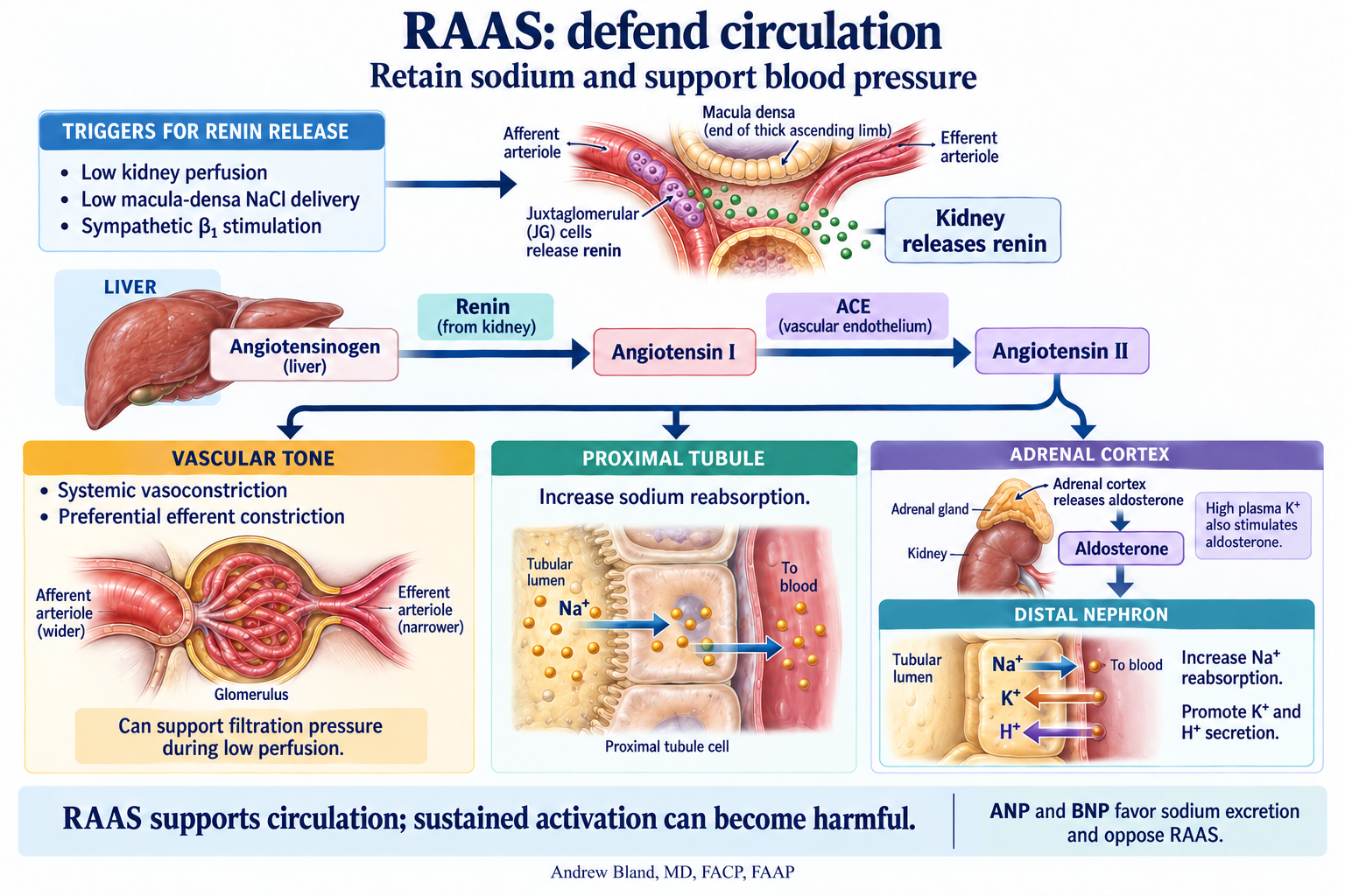
The renin–angiotensin–aldosterone system responds to signals such as reduced kidney perfusion, reduced macula-densa salt delivery, and sympathetic stimulation.
Kidney renin initiates the pathway; angiotensin II supports vascular tone and sodium retention. Adrenal aldosterone promotes distal sodium reabsorption and potassium and hydrogen-ion secretion.
ADH · conserve water
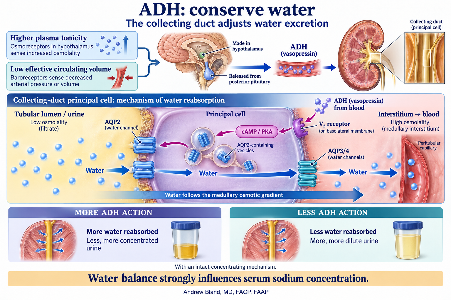
Antidiuretic hormone (vasopressin) is made in the hypothalamus and released from the posterior pituitary. Increased tonicity and reduced effective circulating volume stimulate its release.
ADH increases collecting-duct water permeability through aquaporin-2. This helps explain why a circulation problem can also become a serum sodium problem.
RAAS: from a circulation signal to sodium retention
Trigger and cascade: reduced renal perfusion, reduced macula-densa NaCl delivery, and sympathetic β₁ stimulation promote renin release from juxtaglomerular cells. Renin cleaves liver-derived angiotensinogen to angiotensin I; endothelial ACE forms angiotensin II.
Angiotensin II: supports systemic vascular tone and proximal sodium reabsorption. Preferential efferent constriction can help preserve glomerular pressure during reduced perfusion, but severe hypoperfusion still lowers GFR. Sustained activation can contribute to hypertension and kidney injury.
Aldosterone: comes from the adrenal cortex and promotes distal sodium reabsorption, potassium secretion, and acid secretion. High plasma potassium also directly stimulates its release.
Connect to the laboratory pattern: Why can aldosterone excess combine hypertension, low potassium, and metabolic alkalosis? What changes when an ACE inhibitor or ARB reduces angiotensin II signaling?
ADH: from a water signal to a collecting-duct response
ADH changes water permeability; the medullary osmotic gradient drives water reabsorption. Select the diagram to enlarge it. · Visual library: diagram and teaching text
Trigger and source: increased plasma tonicity stimulates ADH. Low effective circulating volume is another powerful stimulus, even when plasma sodium is low. ADH is made in the hypothalamus and released from the posterior pituitary.
Receptor and channel: ADH binds the basolateral V₂ receptor on collecting-duct principal cells. cAMP/PKA signaling moves AQP2 water channels into the apical, urine-facing membrane. Water exits the cell through basolateral AQP3/4 channels.
Response: with an intact concentrating mechanism, more ADH action means more water reabsorption, less urine volume, and more concentrated urine. Reduced ADH action permits greater excretion of dilute urine. Serum sodium depends on water balance relative to body solute, not simply on how much sodium was eaten.
Connect to the cases: A patient with heart failure can have edema, low effective arterial circulation, persistent ADH release, and hyponatremia at the same time. Which finding describes total-body fluid, and which describes tonicity?
Compare the two systems: RAAS helps defend sodium balance and circulation. ADH controls the collecting duct’s water permeability. They can activate together, but their effects are different.
Other signals that connect the organs
Signal
Where it comes from
Kidney connection
Erythropoietin · EPO
Predominantly kidney interstitial cells, responding to hypoxia.
Stimulates marrow red-cell production. Epogen is a brand of epoetin alfa, a medication that replaces the hormone’s action.
Active vitamin D · calcitriol
Activated in kidney proximal tubules.
Promotes intestinal calcium and phosphate absorption and participates in feedback with PTH.
Natriuretic peptides · ANP and BNP
Heart muscle, responding to stretch.
Favor sodium excretion and counter RAAS-mediated sodium retention. Their effects can be blunted in heart failure, so high peptide levels can coexist with congestion.
Parathyroid hormone · PTH
Parathyroid glands.
Acts on the kidney to conserve calcium, increase phosphate excretion, and stimulate calcitriol production. CKD can disrupt this feedback and raise PTH.
A small set of familiar tests can open the door to many different kidney problems—when we interpret them together.
Start with the basics: blood work, urinalysis, and kidney imaging. The key is the pattern across these findings, interpreted alongside the history, medications, examination, and previous results.
The kidney’s product is homeostasis. Blood tests show the internal environment; urine shows clues to the kidney’s response; imaging adds anatomy and the possibility of obstruction.
Opening question: What finding first brought this patient’s kidneys to your attention?
Creatinine and BUN may bring kidney function to attention. Sodium, potassium, chloride, and bicarbonate reveal water, electrolyte, and acid–base problems. Review calcium, magnesium, and phosphorus when relevant.
Start with the BMP, then compare with the patient’s baseline. A creatinine value needs a date and a trend.
Blood, protein, specific gravity, and the sediment can reveal a problem even when creatinine is normal. Microscopy helps interpret dipstick findings.
Urine albumin-to-creatinine ratio (UACR) quantifies albumin loss. Urine electrolytes and osmolality are additional tests when the clinical question requires them; they are not all part of a routine urinalysis.
Hydronephrosis, stones, cysts, masses, or changes in kidney size may be the first clue. Sometimes the abnormality is found incidentally during imaging for another problem.
Imaging can suggest obstruction or chronic structural disease. Interpret the finding with the history and laboratory results; not every incidental cyst explains impaired function.
Pause and connect: Which finding is the entry point? What would the other two parts of the key add? Topic links open in a new tab so you can return to the lecture.
06 · From a blood result to filtration
Creatinine: a window into kidney function
Serum creatinine (Cr) is a practical blood marker of kidney filtration. It comes largely from muscle creatine metabolism, is freely filtered at the glomerulus, and is also secreted by the proximal tubule. Its blood concentration reflects the balance between production and removal.
GFR is the filtration rate
Glomerular filtration rate describes how much plasma the kidneys filter per minute. With stable creatinine production and handling, a lower GFR generally means a higher serum creatinine once a new steady state is reached. The relationship is inverse and nonlinear.
eGFR is an estimate
Creatinine-based equations combine the blood result with factors such as age and sex to estimate GFR. Muscle mass, diet, and medicines that alter creatinine secretion can affect the estimate. A creatinine inside the laboratory reference range can still accompany reduced filtration.
Read function with context
Compare the baseline and trend. During AKI, creatinine lags behind a changing GFR. Pair filtration with urine albumin and sediment: albuminuria can reveal injury despite a preserved eGFR. GFR alone does not describe every tubular or hormonal kidney function.
Connect the number to the physiology: Blood flow, afferent and efferent arteriolar tone, and filtration pressure influence GFR. Creatinine is a downstream clue to that process; it does not identify the cause by itself.
Start with Normal Physiology, then compare NSAIDs and ARB / ACEi. Which arteriole changes, and what happens to glomerular pressure and GFR?
If creatinine production and handling stay constant, which direction would serum creatinine move after a new steady state is reached? Why would it take time?
Return to albuminuria and macula-densa feedback: how can changing pressure affect both filtration and long-term glomerular stress?
The model illustrates physiologic relationships; its displayed numbers are teaching examples. Interpret a patient’s creatinine change with the full clinical picture.
Albumin/creatinine: pressure, permeability, and risk
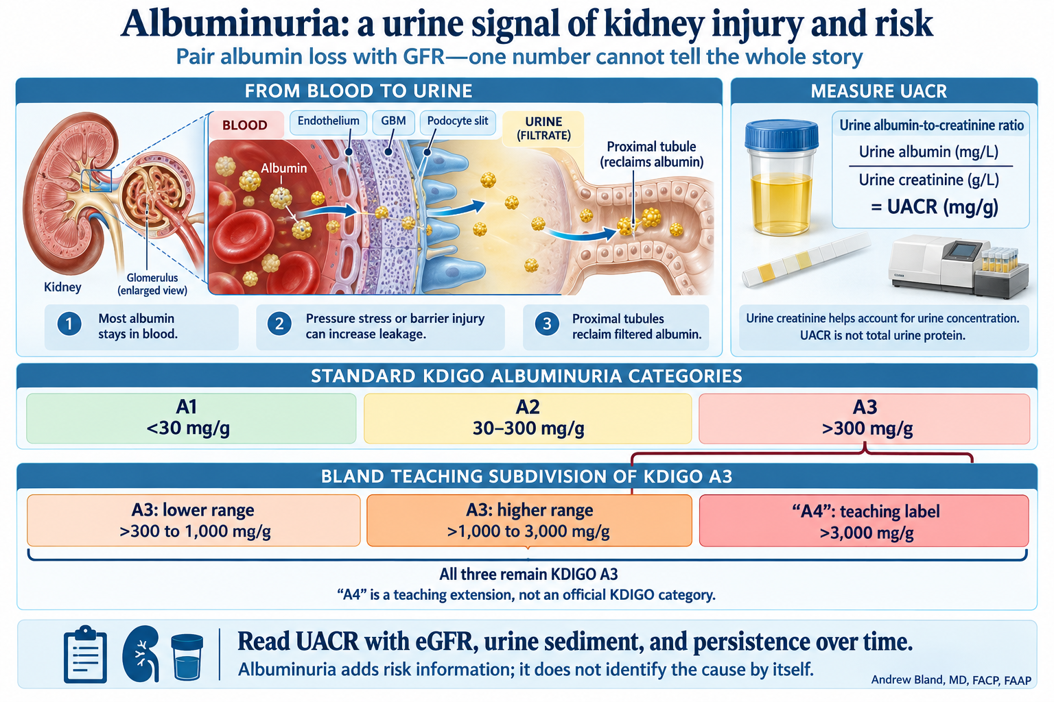
Urine albumin-to-creatinine ratio (UACR, or Alb/Cr) estimates albumin loss while accounting for urine concentration. Albuminuria can be a clue to intraglomerular hypertension and hyperfiltration-related stress, or to inflammatory injury of the glomerular filtration barrier.
Use urine values in the ratio: urine albumin (mg/L) ÷ urine creatinine (g/L) = UACR (mg/g). The urine creatinine helps account for urine concentration; it is not the serum creatinine used to estimate GFR. Differences in creatinine excretion can still influence the ratio.
Three teaching buckets inside the broad A3 category
Bland teaching categories · UACR in mg/g
Teaching label
UACR
Standard KDIGO category
A1
<30
A1
A2
30–300
A2
A3: lower range
>300 to 1,000
A3
A3: higher range
>1,000 to 3,000
A3
“A4”: teaching label
>3,000
A3
The scorecard-inspired teaching extension separates the broad A3 band into three buckets. The upper boundary belongs to the preceding bucket: 1,000 is in the lower A3 range, 3,000 is in the higher A3 range, and values above 3,000 enter the teaching “A4” bucket. “A4” is not an official KDIGO category; all three buckets remain KDIGO A3.
A4 has a published history. The Canadian Society of Nephrology’s 2015 AJKD commentary proposed A4 for nephrotic-range albuminuria (ACR >2,220 mg/g). AJKD is the NKF journal. This lecture uses >3,000 mg/g for its teaching A4; current KDIGO remains A1–A3.
Ask the room: Two people both have persistent eGFR 52, but UACR is 400 in one and 4,000 mg/g in the other. Both are KDIGO G3a A3; what important difference does the broad label hide? The extended view makes the magnitude of albumin loss visible. It does not give an individual risk percentage or diagnose nephrotic syndrome by itself.
Pressure and hyperfiltration
Excess pressure and filtration workload in individual nephrons can promote albumin leakage. A normal or high whole-kidney eGFR does not rule out injury; surviving nephrons may also hyperfilter when total GFR is reduced.
Inflammation and barrier injury
Glomerular inflammation can allow albumin and blood into urine. Use the sediment, clinical presentation, and other testing to determine the cause.
A marker, not a mechanism test: UACR does not directly measure hyperfiltration or prove inflammation. Interpret it with GFR, microscopy, and the timeline. Fever, infection, or exercise may transiently raise it; confirm persistence when assessing CKD.
Albumin is not all urine protein. A low UACR does not exclude excess free light chains. Disproportionately high total urine protein with little albumin can be a clue to a non-albumin protein process.
The macula densa: a salt sensor, not a volume gauge
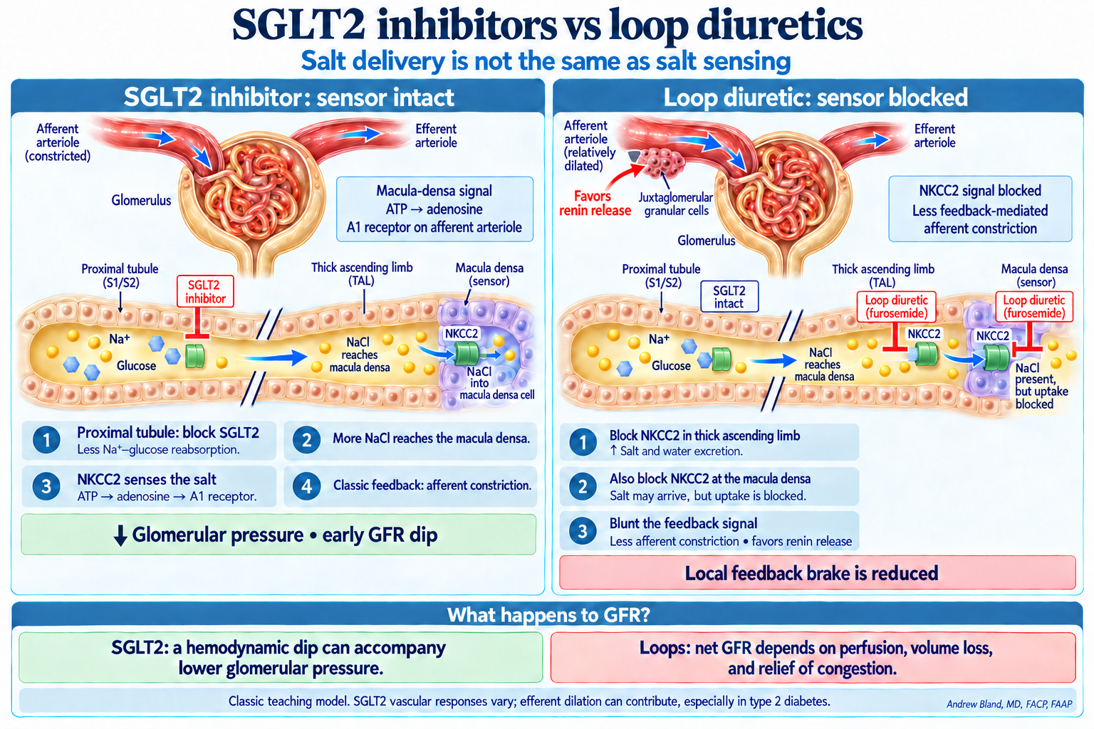
At the end of the thick ascending limb, the macula densa samples tubular NaCl using NKCC2: 1 Na⁺ + 1 K⁺ + 2 Cl⁻. Salt delivery reflects filtration and upstream reabsorption. It provides an indirect signal about perfusion and delivery; it does not directly measure blood volume or serum sodium.
Classic tubuloglomerular feedback: more NaCl sensed → ATP/adenosine signaling → afferent arteriolar constriction → reduced filtration pressure. Low salt delivery promotes renin release and RAAS activation.
Feedback can reduce hyperfiltration and intraglomerular pressure.
Teaching model: restore the signal and apply the afferent “brake.”
Loop diuretic · sensor blocked
Inhibit NKCC2 in the thick ascending limb and macula densa.
Increase salt excretion.
Block the macula densa’s NKCC2 signal despite salt delivery.
Blunt this feedback-mediated afferent constriction and favor renin release.
Teaching model: “blind the sensor.” Volume loss and other signals can still change kidney perfusion and GFR.
Ask the room: if both drugs cause natriuresis, why is the feedback different?
SGLT2 inhibition: delivery increases while the NKCC2 sensor remains functional. A loop diuretic: salt can be present in the lumen while NKCC2-mediated sensing is inhibited. More salt delivered is not the same as more salt sensed.
Then ask: Does a loop diuretic therefore always raise GFR? No. The local feedback brake is reduced, but volume depletion may lower perfusion and GFR; relief of congestion can help kidney function. Interpret the net response in the patient’s clinical context.
Keep the model accurate: SGLT2 responses are not identical in every patient
Afferent constriction is the classic explanation, especially in hyperfiltering type 1 diabetes. Human studies in type 2 diabetes also show responses consistent with efferent vasodilation. The shared concept is reduced intraglomerular pressure; the balance of vascular responses depends on the physiology.
Negatively charged membrane components and binding sites can attract positively charged molecules. In the proximal tubule, binding and receptor-mediated uptake can concentrate some drugs inside cells. Charge helps explain susceptibility; it is one part of a larger mechanism.
Aminoglycosides
These polycationic antibiotics interact with negatively charged sites and the uptake receptor megalin. Proximal-tubule accumulation, including within lysosomes, contributes to cellular injury.
Vancomycin also undergoes tubular uptake, with experimental evidence implicating megalin. Kidney injury depends on exposure, duration, cellular injury pathways, concurrent nephrotoxins, and the patient’s condition.
A simple “less positive, so more is needed” rule does not reliably explain its toxicity or guide dosing.
Monoclonal free light chains—components of antibodies—can injure proximal tubules, form obstructing casts with uromodulin downstream, or contribute to deposits. Protein structure, concentration, and the site of injury matter; positive charge alone does not explain myeloma kidney disease.
Connect back to the key: Tubular injury may appear as rising creatinine, electrolyte losses, or abnormal urine protein. Glomerular albumin leakage and tubular drug uptake involve different structures and mechanisms.
Ask two questions: What was already present? And what has changed now? The same patient can have a chronic kidney problem and a new acute injury.
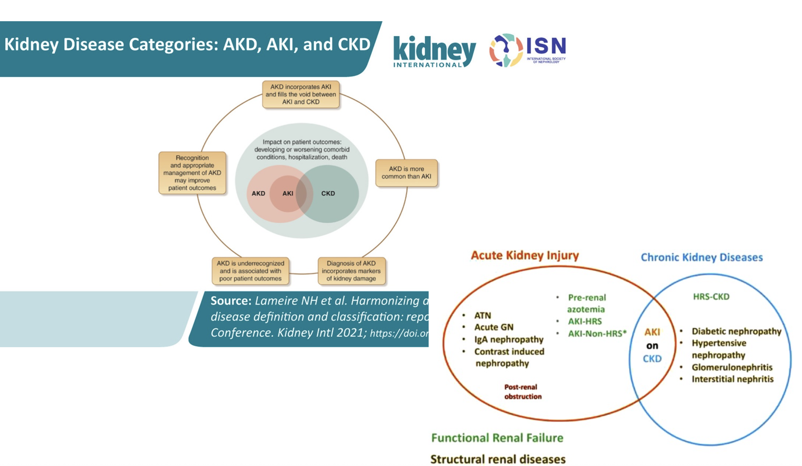
The course AKI–AKD–CKD diagram. Focus on the overlap: AKI on CKD. The disease examples illustrate possible patterns rather than exclusive categories; a disease can have acute and chronic manifestations.
Acute
A new change over hours to days. Compare creatinine with a known baseline and assess urine output.
Chronic
Evidence of abnormal kidney structure or function for at least three months. Prior results matter.
Acute on chronic
A new injury superimposed on established CKD. Describe both the background disease and the current change.
Where does AKD fit? Acute kidney disease is the broader category of kidney abnormalities lasting up to three months and includes AKI. Persistent dysfunction after an acute episode requires reassessment; AKI does not inevitably become CKD.
Discuss the diagram: A patient with a year of reduced kidney function develops vomiting and a new creatinine rise. Why is “AKI on CKD” more informative than choosing only one label?
CKD requires persistent kidney abnormalities for at least three months. Describe the cause, GFR category, and albuminuria category. GFR alone does not tell the whole story.
Move down for lower GFR; move across for greater albuminuria. Both affect risk. Select a cell to discuss its meaning.
The standard view uses KDIGO A1–A3. The teaching view subdivides A3 into >300–1,000, >1,000–3,000, and “A4” >3,000 mg/g. The same KDIGO risk colors apply within A3; these subdivisions do not create additional validated risk classes. Read the historical A4 context.
CKD prognosis categories · GFR in mL/min/1.73 m² · UACR in mg/g
GFR category
A1 <30
A2 30–300
A3 >300
G1 ≥90
G2 60–89
G3a 45–59
G3b 30–44
G4 15–29
G5 <15
Choose any cell. Compare G3a A1 with G3a A3: the same GFR category can carry very different risk.
Green needs context: G1 or G2 with A1 is not CKD unless another marker of kidney damage is present. One abnormal result cannot establish chronicity. This teaching grid shows broad risk groups, not an individual probability of kidney failure.
Discussion: same GFR, different urine
Two patients have a stable eGFR of 52 for six months. One has UACR 12 mg/g; the other, persistently 650 mg/g. They share G3a but fall in A1 and A3. The urine result changes the risk assessment.
Creatinine is a concentration that takes time to respond to a change in filtration. Routine creatinine-based eGFR equations assume a relatively steady state. When creatinine is changing rapidly, the reported eGFR may misrepresent current kidney function.
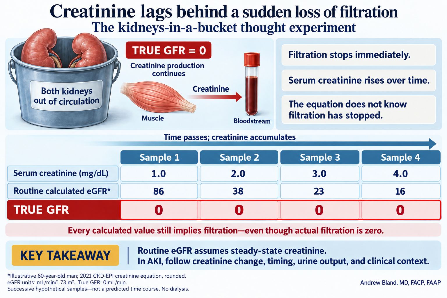
Your kidneys in a bucket: creatinine changes while GFR stays at zero
Imagine taking both kidneys out of the circulation and placing them in a bucket. The patient’s true GFR is immediately zero, but the creatinine already in the blood does not instantly change. Muscle continues producing creatinine, so its concentration rises over time.
The changing creatinine does not require another change in GFR: filtration stays at zero throughout this thought experiment. The routine equation still returns positive numbers because it assumes steady-state creatinine. These values are not all normal, but every one overestimates actual filtration here.
Same patient, four hypothetical samples after filtration stops
Sample
Serum creatinine (mg/dL)
Routine calculated eGFR (mL/min/1.73 m²)
True GFR (mL/min)
1
1.0
86
0
2
2.0
38
0
3
3.0
23
0
4
4.0
16
0
Illustrative 60-year-old man; 2021 CKD-EPI creatinine equation, rounded. These are successive hypothetical samples, not fixed time intervals or a predicted rate of rise. No dialysis is assumed. In real AKI, creatinine production, distribution volume, and residual clearance also affect the trajectory.
Ask the room: When creatinine rises from 2 to 4, did GFR fall again? No—true GFR was already zero. What is changing is the concentration of a substance that continues to accumulate.
Use this in AKI: Follow the creatinine change and timing, urine output, and the clinical picture. Do not treat a routine reported eGFR as current measured filtration or use it to assign a CKD stage during a rapidly changing episode. Specialized kinetic estimates address a different question; they are not the routine laboratory eGFR.
After filtration falls, creatinine accumulates. The reported eGFR can overestimate current filtration while creatinine is still rising.
Creatinine falling
During recovery, creatinine takes time to fall. The reported eGFR can underestimate improving filtration.
Recognize AKI using the change and timing: creatinine rises by ≥0.3 mg/dL within 48 hours, or to ≥1.5 times baseline within the prior seven days; urine output <0.5 mL/kg/hour for six hours is another criterion. AKI can occur with preserved urine output.
Illustrative teaching example · serum creatinine
Two days ago
Today
The important observation
1.0 mg/dL
1.6 mg/dL
A rise of 0.6 mg/dL in 48 hours meets a creatinine criterion for AKI.
Why not assign a CKD stage from today’s eGFR?
The creatinine is not at steady state, and a two-day change does not establish chronic kidney disease. Review the baseline, urine output, volume status, medications, urinalysis, and possible obstruction. Describe the acute trajectory and reassess after stabilization.
Ask before interpreting a number: What was the baseline? When was it measured? Is creatinine rising, falling, or stable?
Both AKI and CKD live in the clinic and the hospital
Pattern
Outpatient encounter
Inpatient encounter
AKI
A new creatinine rise after a diarrheal illness, a medication change, or urinary retention.
A new creatinine rise with sepsis, surgery, or acute heart failure.
CKD
Persistent albuminuria or reduced GFR during longitudinal follow-up.
Established CKD that changes how we interpret an admission and its complications.
AKI on CKD
A patient with CKD develops an acute illness between clinic visits.
The same patient is admitted with an acute change on top of a chronic baseline.
A clinic result may reveal an urgent problem. A hospital patient may have a stable chronic abnormality. The timeline defines the pattern; severity and clinical context determine the urgency.
14 · One patient, several disturbed systems
Expect combinations of disorders
Volume loss + hormonal response
Vomiting can combine chloride loss, metabolic alkalosis, and potassium depletion. Reduced effective circulation activates RAAS and ADH; impaired perfusion may add AKI. The blood and urine reflect both the illness and the kidney’s response.
CKD + several lost functions
Reduced excretory and endocrine reserve can combine hypertension or fluid overload, hyperkalemia, metabolic acidosis, anemia, and altered phosphate–vitamin D–PTH balance. Not every patient has every abnormality.
Proximal injury + multiple urine losses
Some light-chain disorders cause proximal tubular dysfunction: phosphate and bicarbonate wasting, glycosuria despite normal blood glucose, and other losses. The combination suggests a tubular process, sometimes a Fanconi syndrome.
Use physiology to organize the combination: Which nephron function is affected? Which hormones are responding? What is an appropriate compensation, and what represents additional disease?
“We will use cases based on clinical problems I have seen in the real world to show how these nephrology concepts interrelate and overlap.” — Andrew Bland, MD, FACP, FAAP
The cases are fictionalized for teaching. The clinical reasoning is the point: an electrolyte problem may also be a medication problem, a volume problem, and AKI on CKD.
Illness + medications + kidney function
Use the blood results and timeline to connect an acute illness with the chronic background.
Carry these questions into every case: Which kidney jobs and hormonal responses connect the findings? What brought the problem to attention? What is acute, chronic, or both? How do the blood work, urine, and imaging fit together?
Follow these ideas through the case series
The linked topic lectures above are supplementary explanations. The five sessions below organize the assigned case discussions and self-study.
Mechanism studies are linked beside the relevant discussion above. Additional sources for kidney function, hormonal complications, and the AKI–CKD framework:
The overlap image is the existing course teaching graphic. The standard heat-map view renders KDIGO’s conventional GFR–albuminuria risk categories. The optional Bland teaching view subdivides A3 without changing its KDIGO classification or risk colors.