1 · Approach
Use the opening diagram and short lessons before the patient.
AKI, stones, obstruction and calcium
Andrew Bland, MD, FACP, FAAP · October 21, 2026
4 cases now use the same sequence, with 29 short lessons and opening, interpretation and treatment figures.
Open the collected self-study teaching ↓ · Browse the self-study case guide
Opening frameworks, 35 Learn first sections, stage-specific infographics and direct lecture links for the three classroom cases. Use the short lesson before its question, then return to the case to commit, discuss and reveal.
Session plan and self-study cases · Shared opening lecture · Day 4 figures in the visual library
Look for severe electrolyte disturbance, shock, rhabdomyolysis, tumor lysis and an infected obstructed kidney. Treat the threat while deciding whether AKI is prerenal, intrinsic, postrenal or mixed.
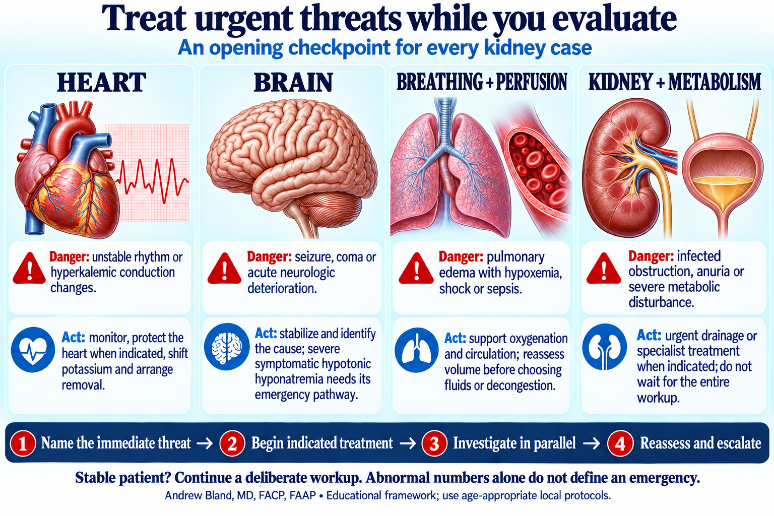
Name the threat, begin indicated treatment, investigate in parallel, then reassess.
Start with perfusion, urine and imaging; compare ischemic tubular injury with pigment injury and track the accompanying electrolyte risks.
Use the opening diagram and short lessons before the patient.
Revisit urgency as history, examination and initial data arrive.
Use the urine, blood or imaging guide at the relevant decision.
Connect the management plan with response, monitoring and follow-up.
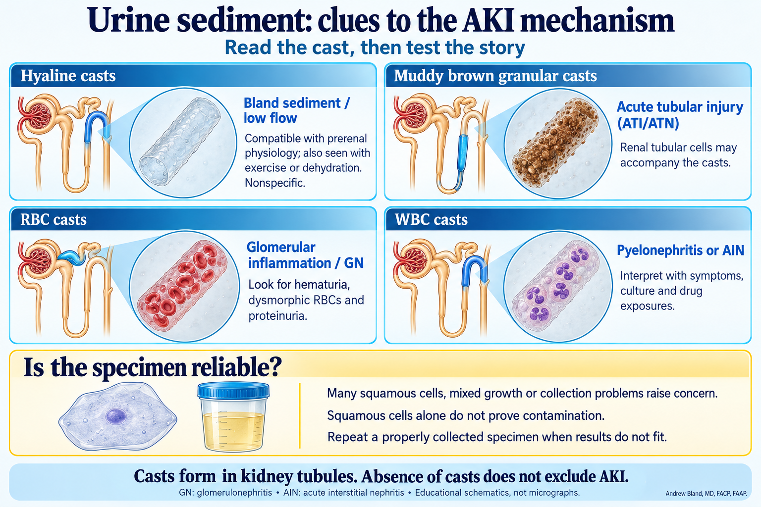
Use the diagram with the teaching text, then apply it to the case findings.
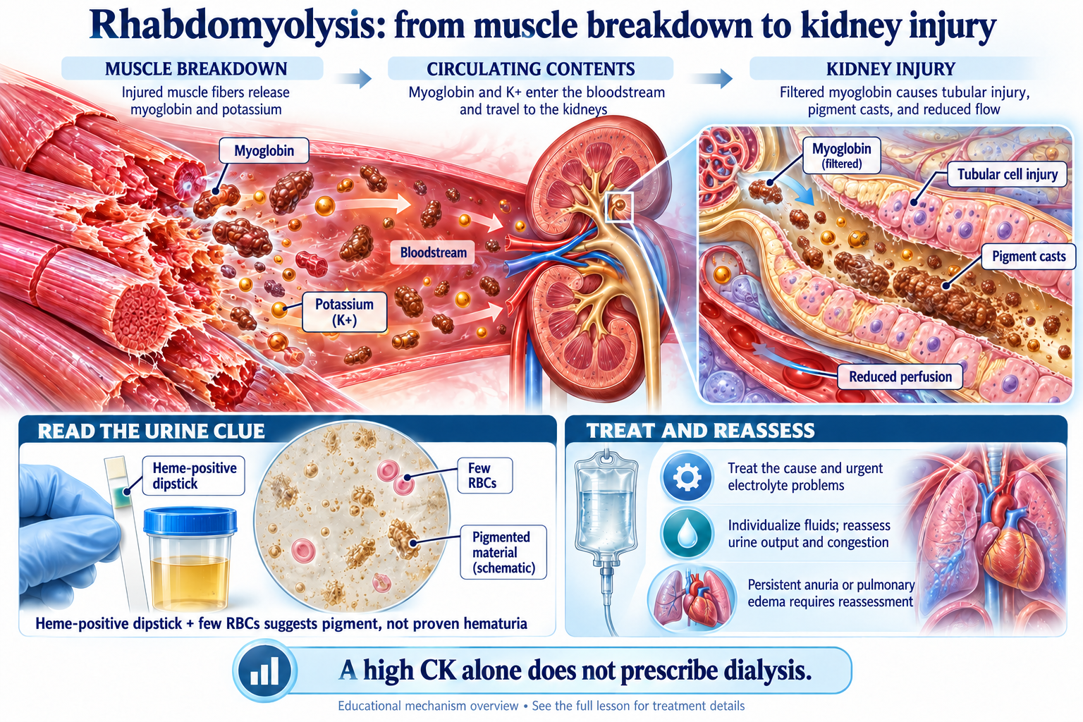
Use the diagram with the teaching text, then apply it to the case findings.
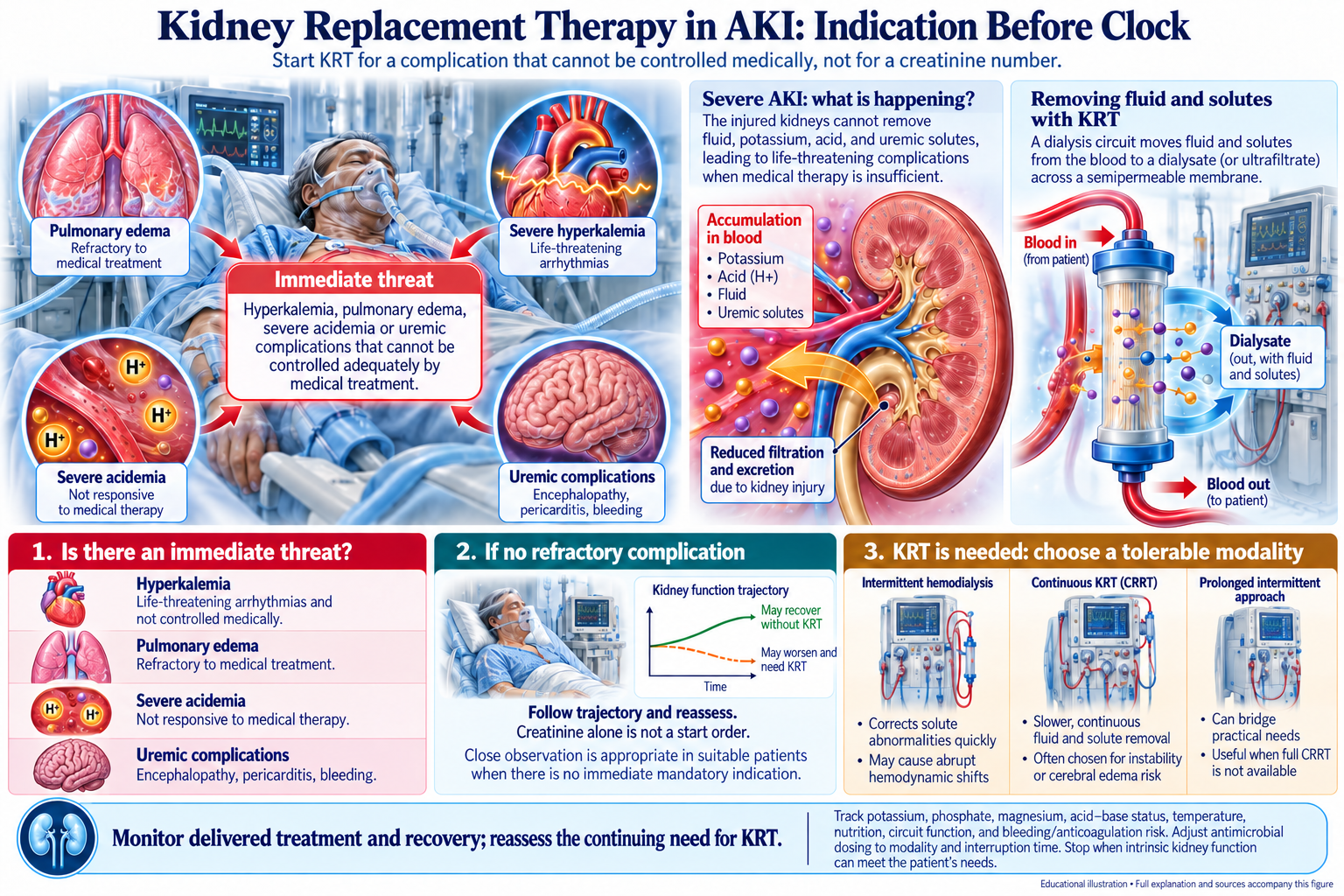
Use the diagram with the teaching text, then apply it to the case findings.
Assigned reading: the explanations in this case and the question-linked course evidence guide. The objective map distinguishes tested decisions from concepts taught in explanations.
Concepts: aki-01, aki-02, aki-03, aki-04, aki-05, aki-06, aki-08, aki-09, aki-11, aki-12, aki-13, aki-15, aki-22, aki-23, aki-24, aki-25, glom-01, lytes-17, lytes-19, lytes-21.
Rhabdomyolysis lecture — Clinical Diagnosis & Emergency Management
Renal pharmacology lecture — Drug Dosing: Principles of Renal Clearance
Find the level of obstruction, identify whether infection or a solitary kidney changes urgency, and anticipate a different risk after drainage.
Use the opening diagram and short lessons before the patient.
Revisit urgency as history, examination and initial data arrive.
Use the urine, blood or imaging guide at the relevant decision.
Connect the management plan with response, monitoring and follow-up.
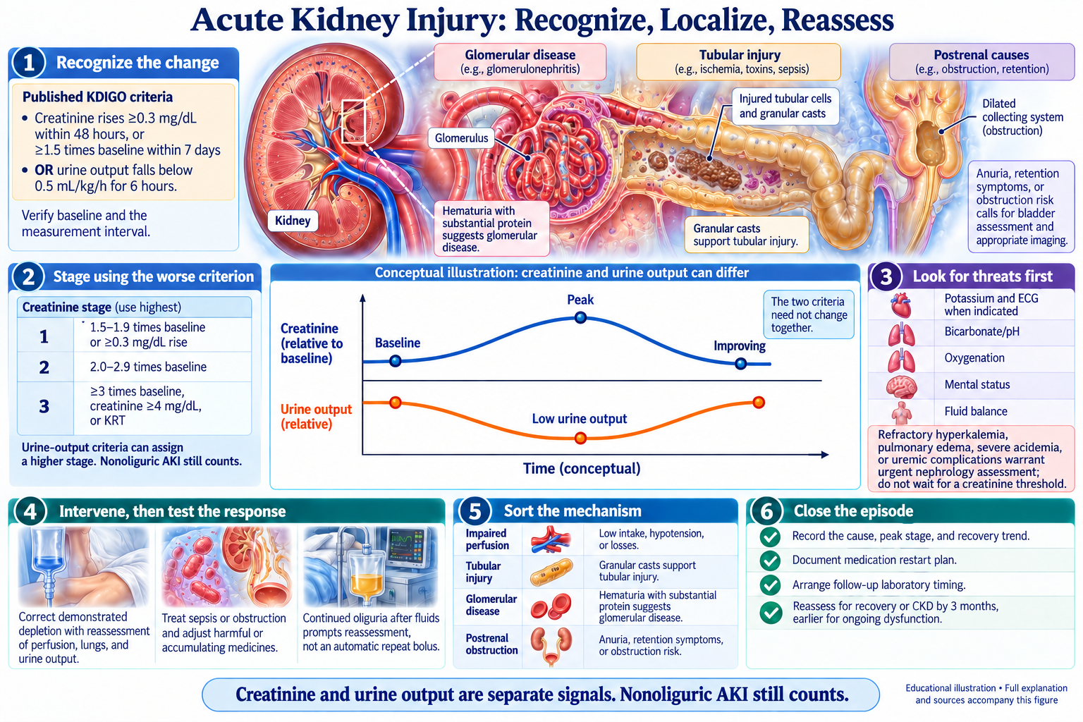
Use the diagram with the teaching text, then apply it to the case findings.
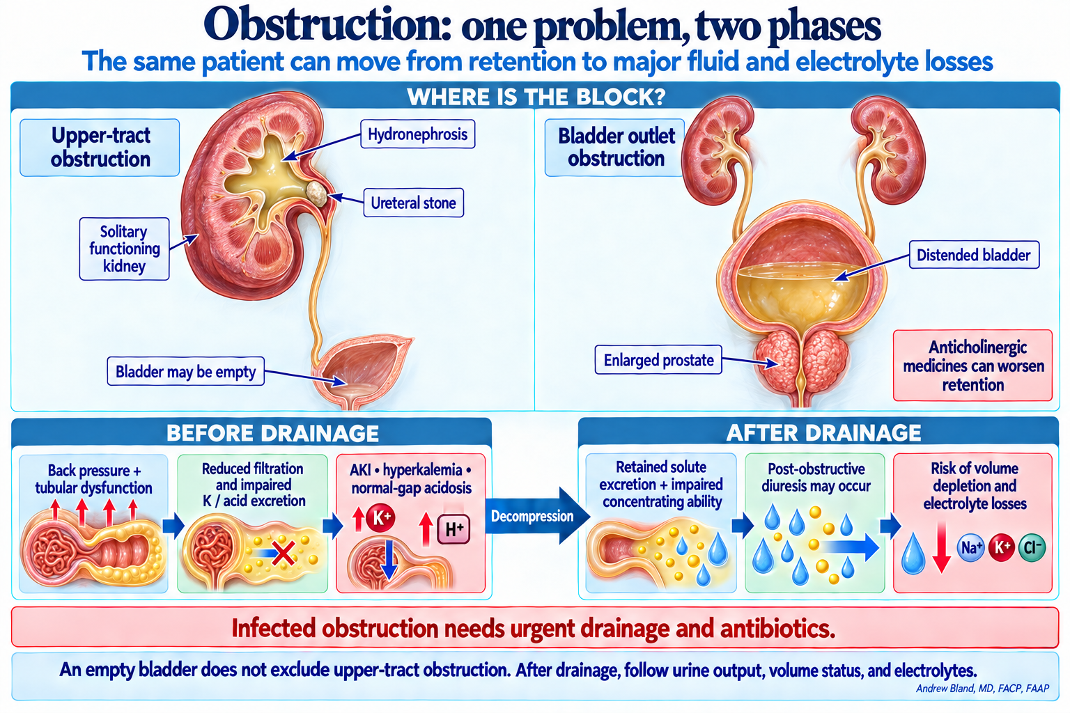
Use the diagram with the teaching text, then apply it to the case findings.
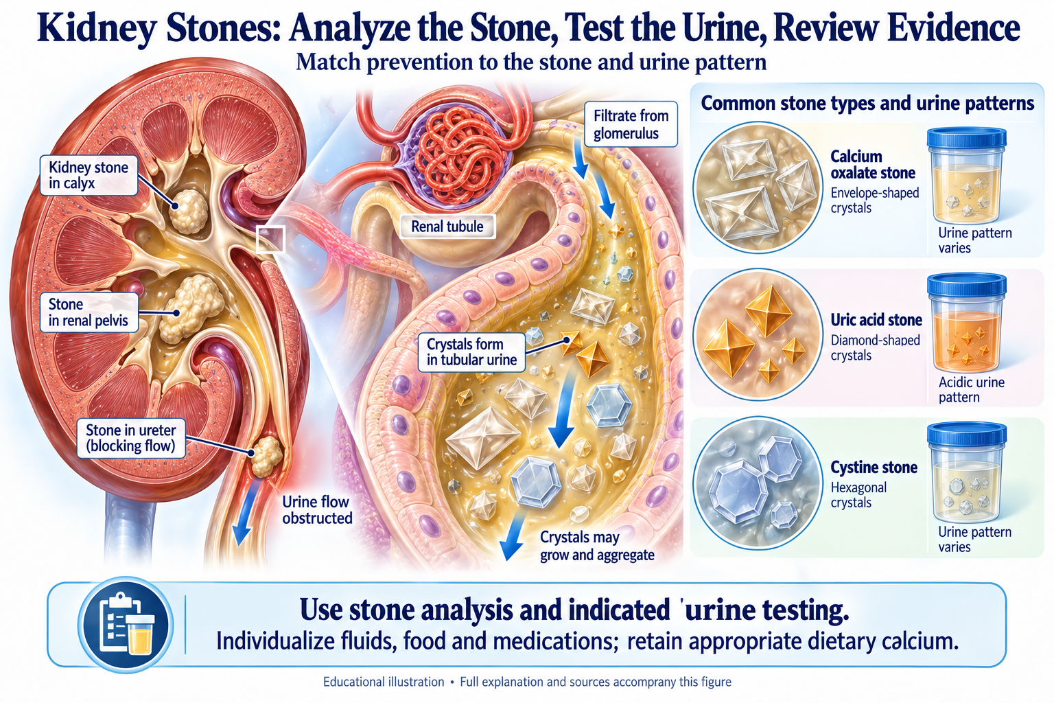
Use the diagram with the teaching text, then apply it to the case findings.
Assigned reading: the explanations in this case and the question-linked course evidence guide. The objective map distinguishes tested decisions from concepts taught in explanations.
Concepts: aki-03, aki-07, aki-13, aki-17, aki-18, aki-22, aki-25, lytes-12, lytes-13, structural-01, structural-02, structural-03, structural-05, structural-06, structural-07, structural-08, structural-14, structural-20.
AKI BMP pattern recognition guide — Part 1: Three-compartment AKI framework
Obstructive uropathy handout — III. Hydronephrosis and upper urinary tract obstruction
Obstructive uropathy handout — IV. AKI without hydronephrosis (NDOU)
Dialysis prescription and complications — Urgent dialysis indications: AEIOU
Hyperkalemia lecture — Recognize risk before choosing a treatment
Hyperkalemia lecture — Protect the myocardium / Shift / Arrange removal
Nephrolithiasis handout — III. Clinical presentation and diagnosis
Nephrolithiasis handout — IV. Medical management / VI. Surgical indications
Nephrolithiasis lecture — Stone types and directed therapy; The metabolic workup
UTI assessment and treatment — Evidence-based UTI treatment guidelines
Obstructive uropathy handout — I. BPH and lower urinary tract obstruction
Obstructive nephropathy imaging review — 2. Renal ultrasound: capabilities and limitations
Renal labs and imaging handout — Section 3: Renal imaging modalities
Confirm severe hypercalcemia, let PTH organize the workup, and evaluate the renal mass without assuming it explains every finding.
Use the opening diagram and short lessons before the patient.
Revisit urgency as history, examination and initial data arrive.
Use the urine, blood or imaging guide at the relevant decision.
Connect the management plan with response, monitoring and follow-up.
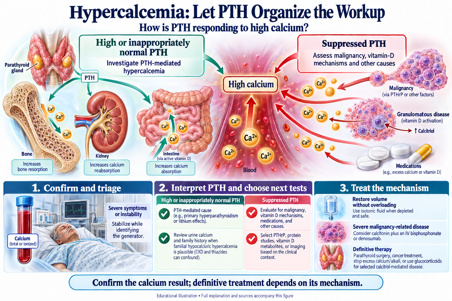
Use the diagram with the teaching text, then apply it to the case findings.
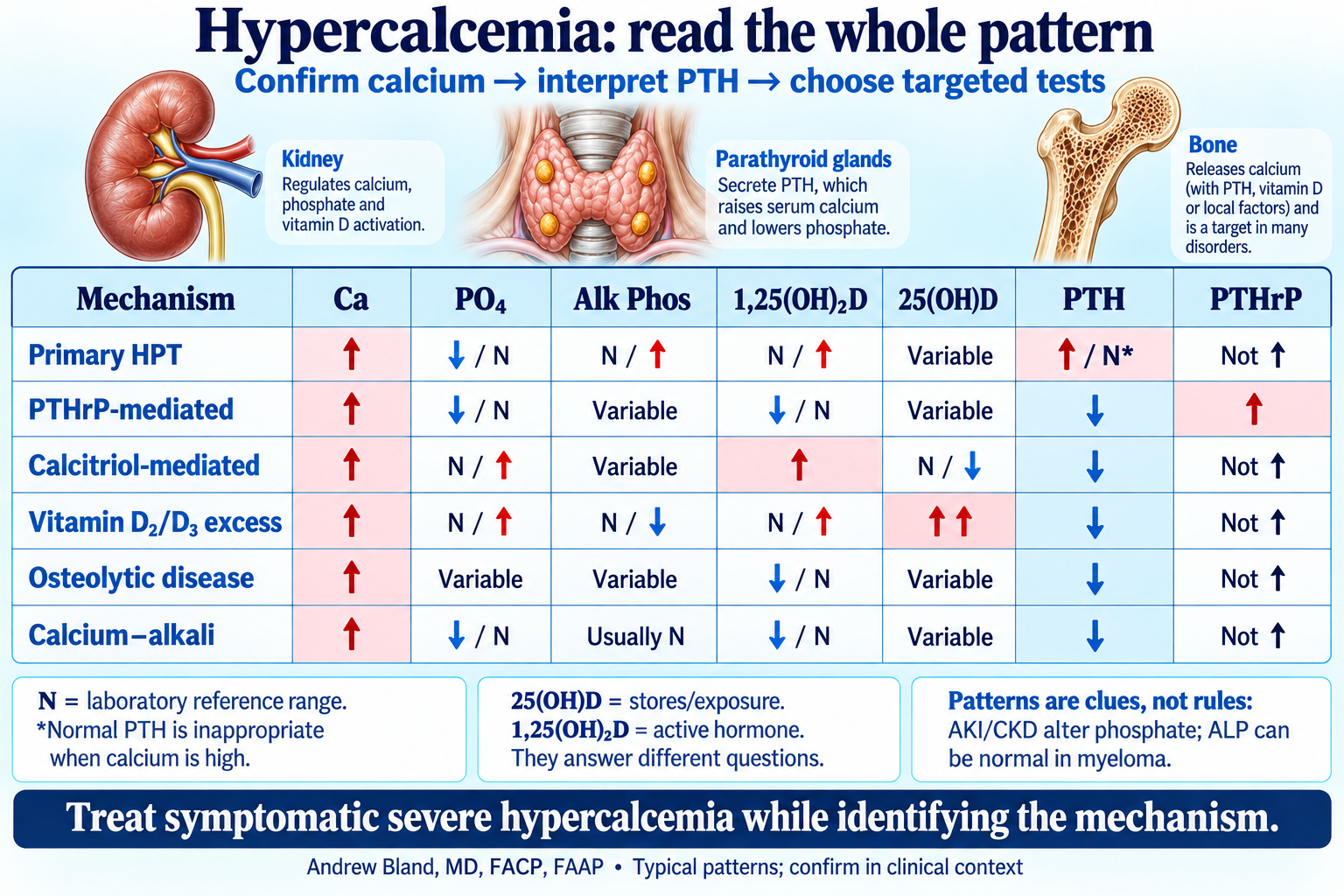
Use the diagram with the teaching text, then apply it to the case findings.
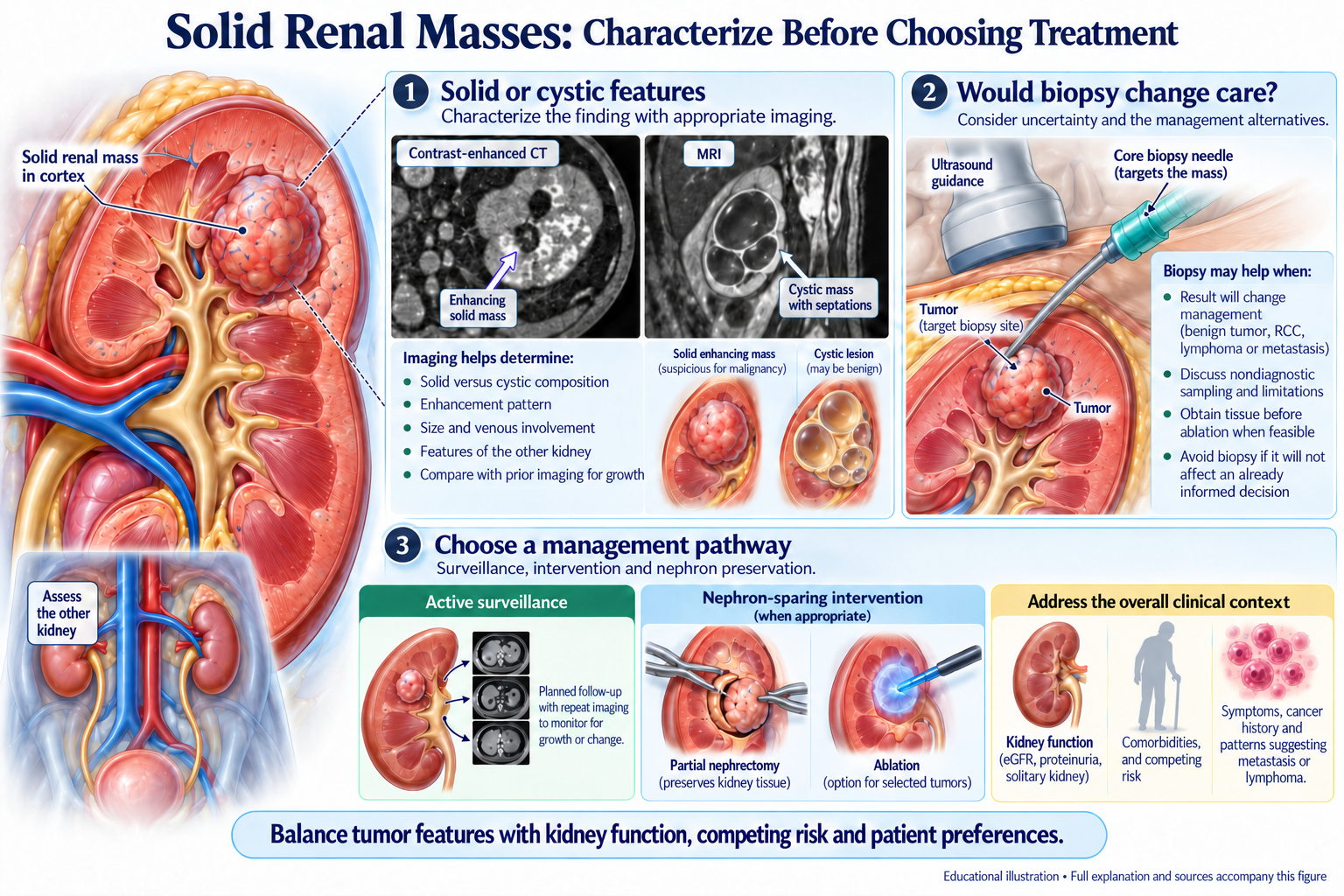
Use the diagram with the teaching text, then apply it to the case findings.
Assigned reading: the explanations in this case and the question-linked course evidence guide. The objective map distinguishes tested decisions from concepts taught in explanations.
Concepts: aki-03, aki-04, aki-07, aki-10, aki-26, aki-27, glom-06, lytes-10, lytes-15, lytes-16, structural-12, structural-13, structural-14, structural-15, structural-23.
Contrast-associated AKI lecture — Prevention; What Does NOT Work
AKI lecture — Post-AKI Follow-Up Care · AKI staging student handout — AKI → CKD Transition: Critical Window
Hypernatremia lecture — Central vs Nephrogenic Diabetes Insipidus
Hypercalcemia lecture — Diagnostic Approach: PTH-Dependent vs PTH-Independent
Renal labs and imaging student handout — Section 3: Renal Imaging Modalities
Renal labs and imaging student handout — Section 4: Contrast Considerations
Drug dosing quick reference — Part 5: Antidiabetic Medications
Bring the lecture to the bedside · Course evidence guide · Integrated case summaries
Open a case below for its teaching, diagrams and lecture links. Use the staged case to answer the questions and reveal explanations.
Map exposures against the creatinine and sediment timeline. Allow overlapping tubular and interstitial injury, and reassess dosing as kidney function changes.
Read the opening framework and urgent-threat checkpoint.
Build the pattern as the history, examination and results arrive.
Use the mechanism to explain the blood, urine or imaging findings.
Connect the treatment with the response, risks and follow-up.
Review the shared urgent-threat framework →
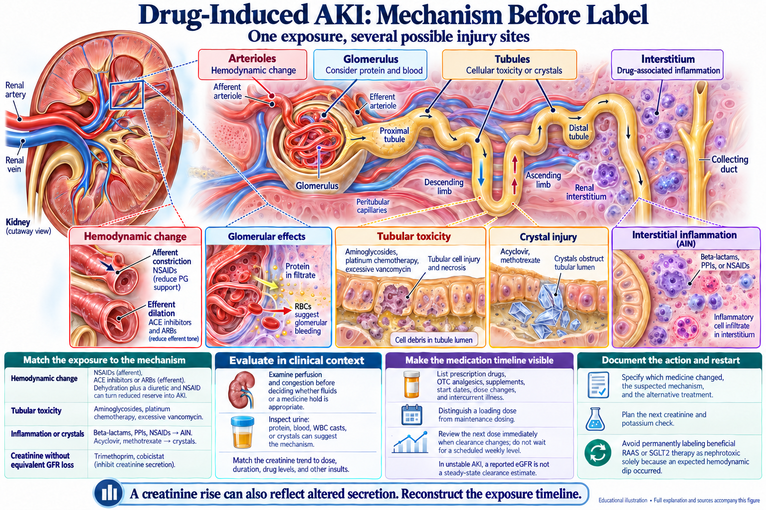
Use the visual framework with the case findings and the linked lecture.
Use the visual framework with the case findings and the linked lecture.
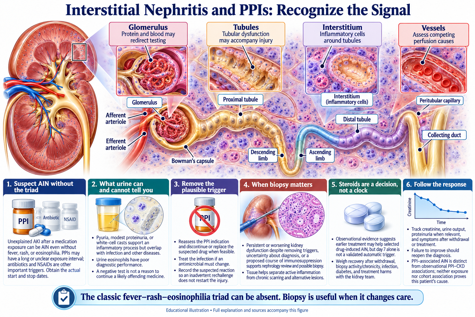
Use the visual framework with the case findings and the linked lecture.
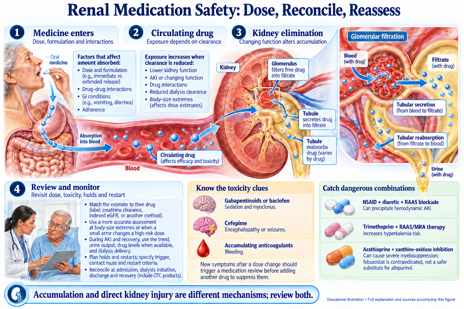
Use the visual framework with the case findings and the linked lecture.
Which findings support more than one AKI mechanism, and how will the medication plan change during recovery?
Assigned reading: the explanations in this case and the question-linked course evidence guide. The objective map distinguishes tested decisions from concepts taught in explanations.
Concepts: aki-02, aki-06, aki-09, aki-10, aki-12, aki-25, aki-26, ckd-18, structural-15, structural-21, structural-23.
Contrast-associated AKI lecture — Prevention; What Does NOT Work
Renal pharmacology lecture — Antibiotic Dosing in AKI and CKD
Renal labs and imaging handout — Section 4: Contrast Considerations
Renal pharmacology lecture — Antibiotic Dosing in AKI and CKD
Drug dosing quick reference — Part 5: Antidiabetic Medications
Drug dosing quick reference — Part 1: Principles of Drug Dosing in CKD
Recognize tumor lysis and treat cardiac, calcium and volume threats in parallel. Review superimposed crystal injury and arrange definitive removal when needed.
Read the opening framework and urgent-threat checkpoint.
Build the pattern as the history, examination and results arrive.
Use the mechanism to explain the blood, urine or imaging findings.
Connect the treatment with the response, risks and follow-up.
Review the shared urgent-threat framework →
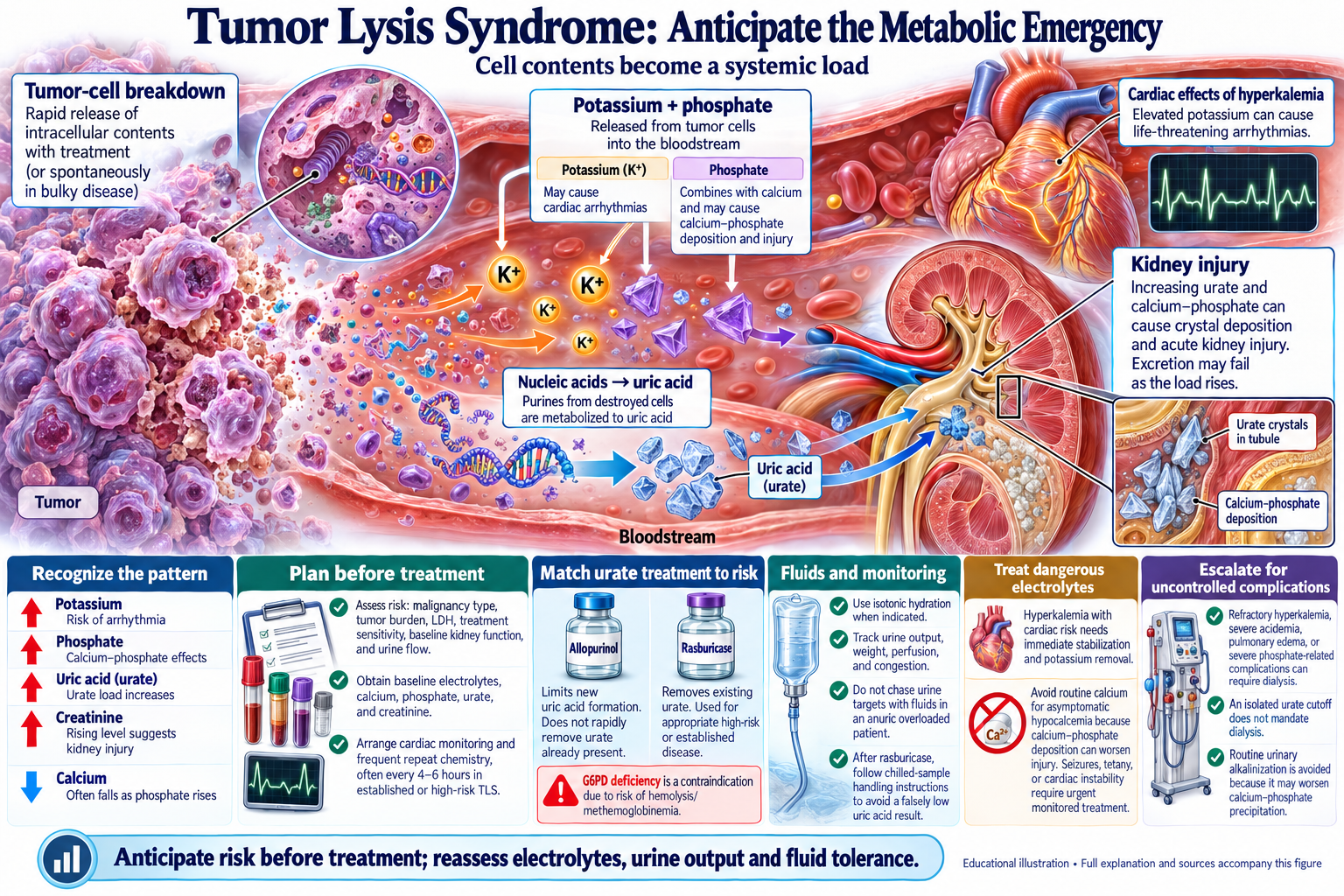
Use the visual framework with the case findings and the linked lecture.
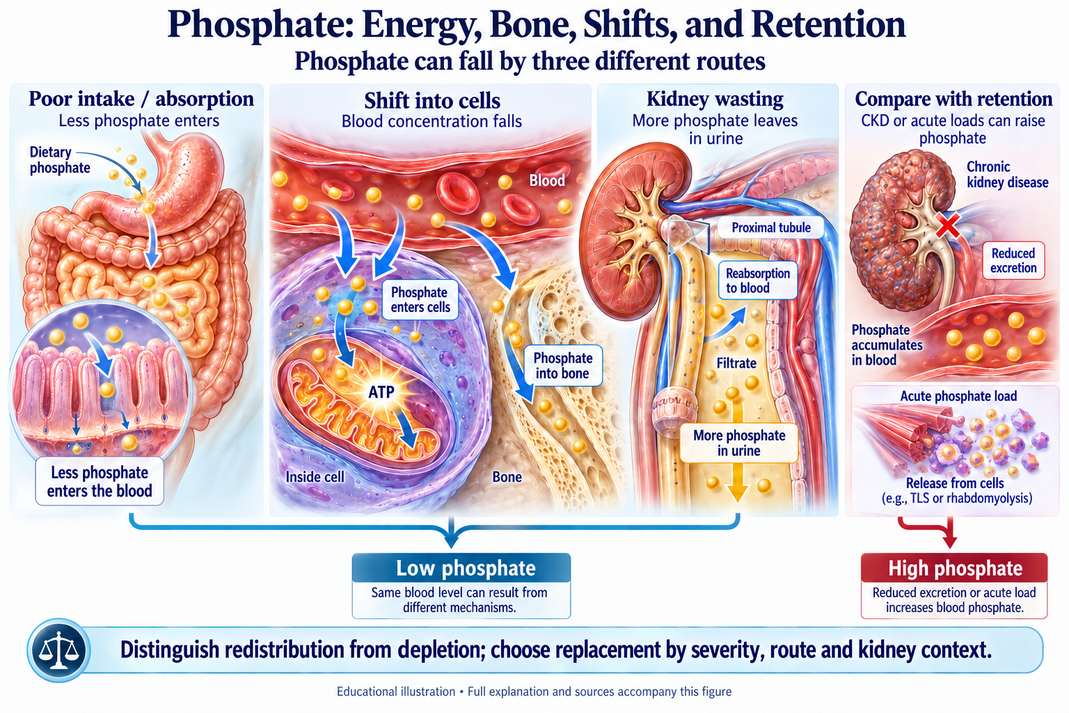
Use the visual framework with the case findings and the linked lecture.
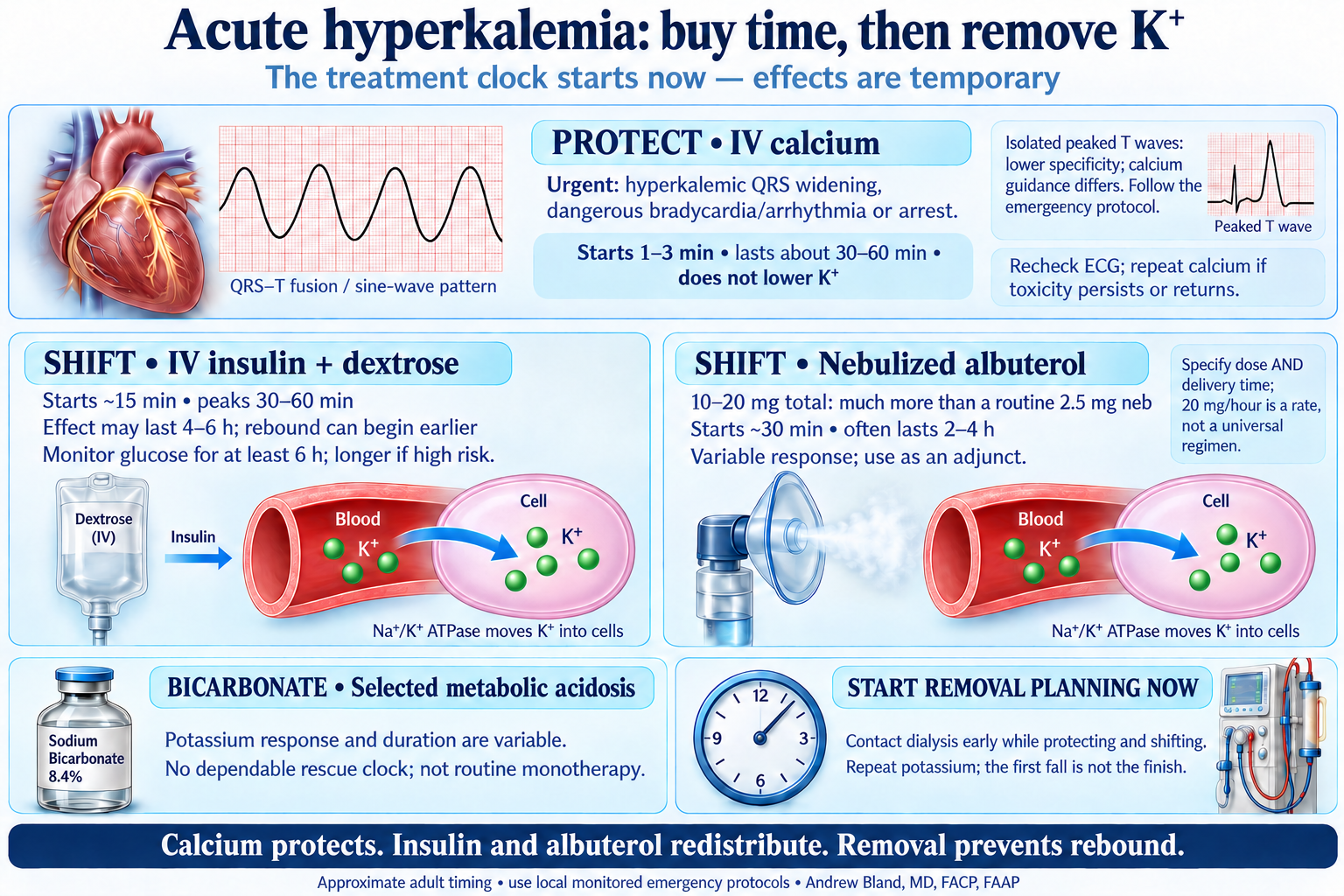
Use the visual framework with the case findings and the linked lecture.
Use the visual framework with the case findings and the linked lecture.
Which treatments stabilize, which shift, which remove, and what continues to drive rebound?
Assigned reading: the explanations in this case and the question-linked course evidence guide. The objective map distinguishes tested decisions from concepts taught in explanations.
Concepts: aki-16, aki-21, aki-22, aki-23, aki-24, aki-25, lytes-13, lytes-17, lytes-19.
AKI lecture — Crystalopathy: Crystal-Induced Tubular Obstruction & AKI · Drug-induced AKI overview — Crystal Nephropathy
TLS lecture — Definition & Pathophysiology; Prevention Strategies
Hypocalcemia lecture — Emergency Protocol: Symptomatic Hypocalcemia
Separate total-body fluid excess from effective arterial filling. Investigate infection, depletion, drugs and obstruction before assigning the HRS phenotype.
Read the opening framework and urgent-threat checkpoint.
Build the pattern as the history, examination and results arrive.
Use the mechanism to explain the blood, urine or imaging findings.
Connect the treatment with the response, risks and follow-up.
Review the shared urgent-threat framework →
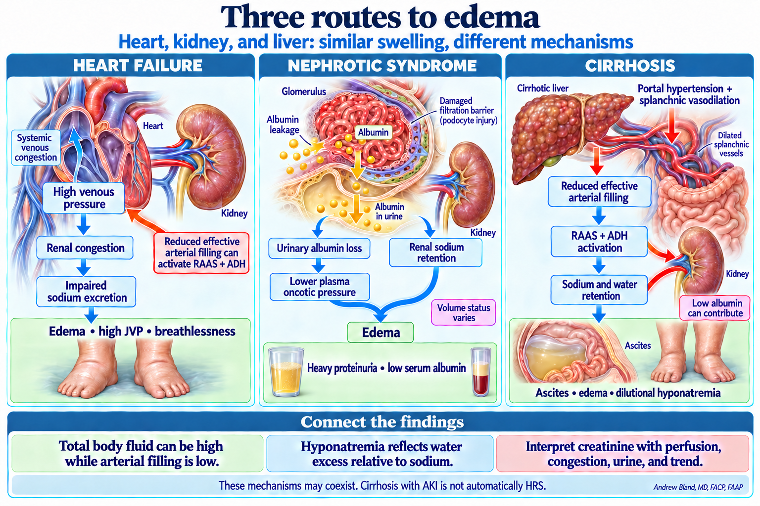
Use the visual framework with the case findings and the linked lecture.
Use the visual framework with the case findings and the linked lecture.
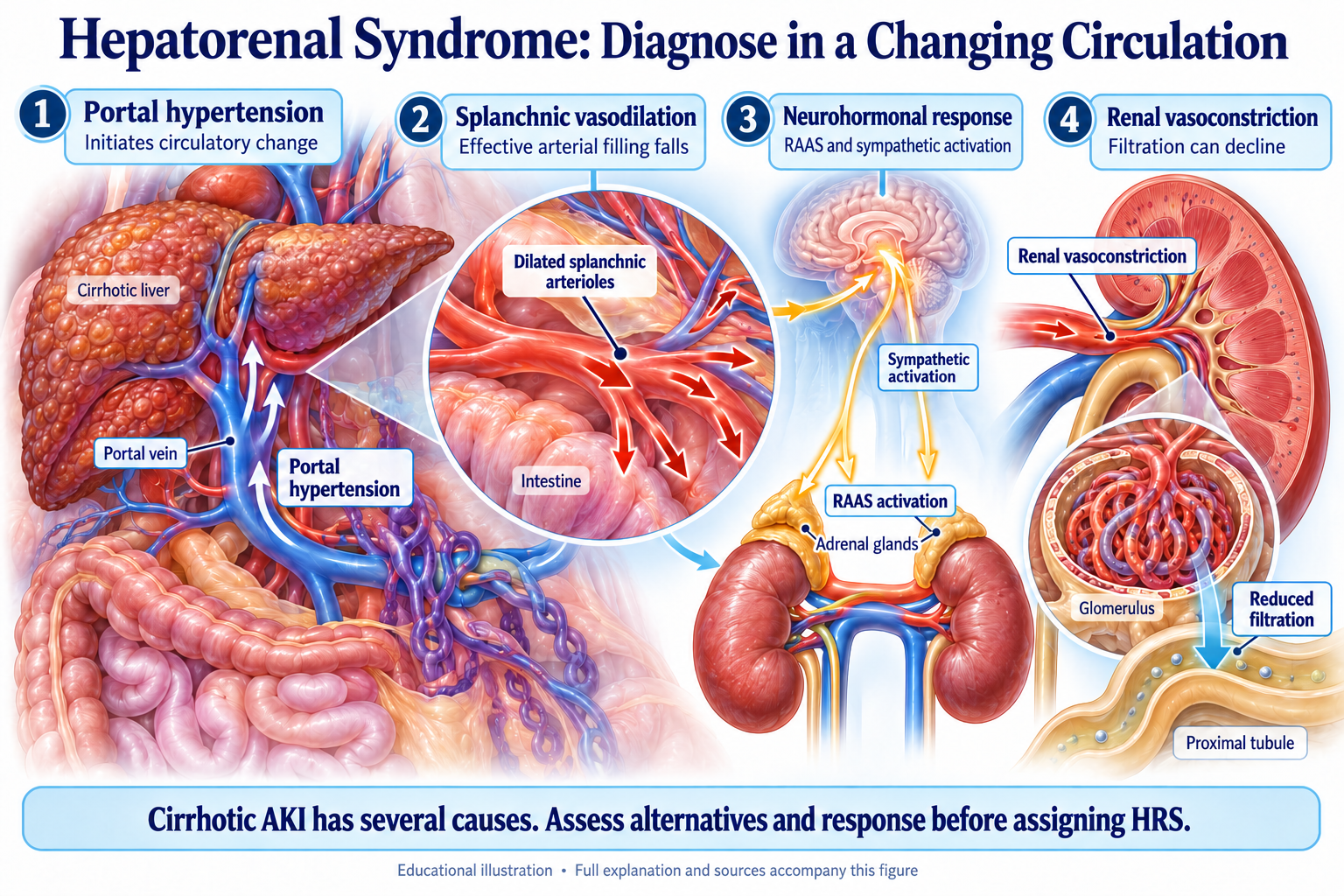
Use the visual framework with the case findings and the linked lecture.
Why does a low urine sodium not establish HRS, and what response to treatment changes the working diagnosis?
Assigned reading: the explanations in this case and the question-linked course evidence guide. The objective map distinguishes tested decisions from concepts taught in explanations.
Concepts: aki-03, aki-04, aki-05, aki-20, aki-23, htn-cardiorenal-26, lytes-07, lytes-14, structural-20.
AKI BMP pattern guide — Part 1: Three-Compartment AKI Framework · AKI lecture — Systematic Diagnostic Approach
AKI lecture — FeNa and FeUrea: Know Their Limits · Urinalysis lecture — FeNa Calculator with Clinical Context
HRS handout — Diagnostic Approach; Management: The Stepwise Approach
HRS handout — Updated Classification: HRS-AKI and HRS-non-AKI; Summary Table: HRS at a Glance
Hyponatremia lecture — Hypervolemic hyponatremia: cirrhosis with ascites
Hyperkalemia lecture — Find the mechanism and reversible contributors · RTA types — Type 4: Hyperkalemic Renal Tubular Acidosis
NSAID-induced AKI lecture — Risk Factors for NSAID-Induced Kidney Injury · Renal pharmacology — 6. NSAID Alternatives for Pain Management in CKD
Identify impaired emptying, relieve significant obstruction and anticipate the change from retention to post-obstructive water and electrolyte losses.
Read the opening framework and urgent-threat checkpoint.
Build the pattern as the history, examination and results arrive.
Use the mechanism to explain the blood, urine or imaging findings.
Connect the treatment with the response, risks and follow-up.
Review the shared urgent-threat framework →
Use the visual framework with the case findings and the linked lecture.
Use the visual framework with the case findings and the linked lecture.
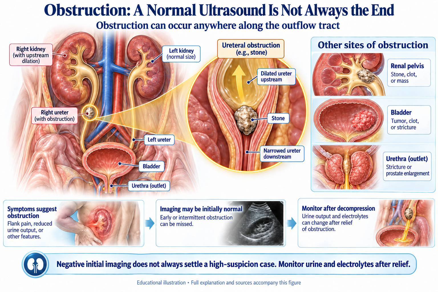
Use the visual framework with the case findings and the linked lecture.
What changes after drainage, and which follow-up decisions prevent recurrent retention and treatment-related falls?
Assigned reading: the explanations in this case and the question-linked course evidence guide. The objective map distinguishes tested decisions from concepts taught in explanations.
Concepts: aki-01, aki-07, aki-17, aki-18, lytes-13, lytes-14, lytes-23, lytes-24, structural-04, structural-06, structural-08, structural-18, structural-25.
Obstructive uropathy handout — III. Hydronephrosis and Upper Urinary Tract Obstruction · Obstructive nephropathy imaging review — 2. Renal Ultrasound: Capabilities and Limitations
Obstructive uropathy handout — III. Hydronephrosis; IV. AKI Without Hydronephrosis (NDOU)
Obstructive uropathy handout — V. Post-Obstructive Diuresis (POD)
UTI assessment lecture — Asymptomatic Bacteriuria; Evidence-Based UTI Assessment Algorithm · UTI prevention handout — Section 8: When to Refer to Urology
Obstructive uropathy handout — V. Post-Obstructive Diuresis (POD)
Geriatric nephrology handout — II. eGFR Interpretation in Elderly; III. Geriatric Drug Dosing in CKD
Hyperkalemia lecture — Find the mechanism and reversible contributors · RTA types — Type 4: Hyperkalemic Renal Tubular Acidosis
Metabolic acidosis lecture — Urinary Anion Gap · Acid-base student guide — 7. Non-Anion Gap Acidosis